Module 26: The Muscular System

Lesson 6: Muscles of the Pelvic Girdle and Lower Limbs

Các Cơ Của Đai Chậu Và Chi Dưới

Nội dung bài học:
Mỗi bài học (lesson) bao gồm 4 phần chính: Thuật ngữ, Luyện Đọc, Luyện Nghe, và Bàn Luận.
Sử dụng tính năng:
Bôi hoặc nhấp đôi vào từ, sau đó ấn vào biểu tượng để tra nghĩa từ đó. Khi bạn đưa chuột đến câu (hoặc khi nhấp vào câu trên màn hình điện thoại), gợi ý về cách hiểu câu đó sẽ hiện lên. Cuối cùng, bạn có thể đánh dấu hoàn thành toàn bộ bài học bằng cách ấn vào nút “Hoàn Thành” ở cuối trang.
Đăng ký và đăng nhập
Bạn cần đăng ký và đăng nhập vào tài khoản để lưu quá trình học.
Dưới đây là danh sách những thuật ngữ Y khoa của module The Muscular System.
Khái quát được số lượng thuật ngữ sẽ xuất hiện trong bài đọc và nghe sẽ giúp bạn thoải mái tiêu thụ nội dung hơn. Sau khi hoàn thành nội dung đọc và nghe, bạn hãy quay lại đây và luyện tập (practice) để quen dần các thuật ngữ này. Đừng ép bản thân phải nhớ các thuật ngữ này vội vì bạn sẽ gặp và ôn lại danh sách này trong những bài học (lesson) khác của cùng một module.

Medical Terminology: The Muscular System

abduct
move away from midline in the sagittal plane
abductor
moves the bone away from the midline
abductor digiti minimi
muscle that abducts the little finger
abductor pollicis brevis
muscle that abducts the thumb
abductor pollicis longus
muscle that inserts into the first metacarpal
adductor
moves the bone toward the midline
adductor brevis
muscle that adducts and medially rotates the thigh
adductor longus
muscle that adducts, medially rotates, and flexes the thigh
adductor magnus
muscle with an anterior fascicle that adducts, medially rotates and flexes the thigh, and a posterior fascicle that assists in thigh extension
adductor pollicis
muscle that adducts the thumb
agonist
(also, prime mover) muscle whose contraction is responsible for producing a particular motion
anal triangle
posterior triangle of the perineum that includes the anus
anconeus
small muscle on the lateral posterior elbow that extends the forearm
antagonist
muscle that opposes the action of an agonist
anterior compartment of the arm
(anterior flexor compartment of the arm) the biceps brachii, brachialis, brachioradialis, and their associated blood vessels and nerves
anterior compartment of the forearm
(anterior flexor compartment of the forearm) deep and superficial muscles that originate on the humerus and insert into the hand
anterior compartment of the leg
region that includes muscles that dorsiflex the foot
anterior compartment of the thigh
region that includes muscles that flex the thigh and extend the leg
anterior scalene
a muscle anterior to the middle scalene
appendicular
of the arms and legs
axial
of the trunk and head
belly
bulky central body of a muscle
bi
two
biceps brachii
two-headed muscle that crosses the shoulder and elbow joints to flex the forearm while assisting in supinating it and flexing the arm at the shoulder
biceps femoris
hamstring muscle
bipennate
pennate muscle that has fascicles that are located on both sides of the tendon
brachialis
muscle deep to the biceps brachii that provides power in flexing the forearm.
brachioradialis
muscle that can flex the forearm quickly or help lift a load slowly
brevis
short
buccinator
muscle that compresses the cheek
calcaneal tendon
(also, Achilles tendon) strong tendon that inserts into the calcaneal bone of the ankle
caval opening
opening in the diaphragm that allows the inferior vena cava to pass through; foramen for the vena cava
circular
(also, sphincter) fascicles that are concentrically arranged around an opening
compressor urethrae
deep perineal muscle in females
convergent
fascicles that extend over a broad area and converge on a common attachment site
coracobrachialis
muscle that flexes and adducts the arm
corrugator supercilii
prime mover of the eyebrows
deep anterior compartment
flexor pollicis longus, flexor digitorum profundus, and their associated blood vessels and nerves
deep posterior compartment of the forearm
(deep posterior extensor compartment of the forearm) the abductor pollicis longus, extensor pollicis brevis, extensor pollicis longus, extensor indicis, and their associated blood vessels and nerves
deep transverse perineal
deep perineal muscle in males
deglutition
swallowing
deltoid
shoulder muscle that abducts the arm as well as flexes and medially rotates it, and extends and laterally rotates it
diaphragm
skeletal muscle that separates the thoracic and abdominal cavities and is dome-shaped at rest
digastric
muscle that has anterior and posterior bellies and elevates the hyoid bone and larynx when one swallows; it also depresses the mandible
dorsal group
region that includes the extensor digitorum brevis
dorsal interossei
muscles that abduct and flex the three middle fingers at the metacarpophalangeal joints and extend them at the interphalangeal joints
epicranial aponeurosis
(also, galea aponeurosis) flat broad tendon that connects the frontalis and occipitalis
erector spinae group
large muscle mass of the back; primary extensor of the vertebral column
extensor
muscle that increases the angle at the joint
extensor carpi radialis brevis
muscle that extends and abducts the hand at the wrist
extensor carpi ulnaris
muscle that extends and adducts the hand
extensor digiti minimi
muscle that extends the little finger
extensor digitorum
muscle that extends the hand at the wrist and the phalanges
extensor digitorum brevis
muscle that extends the toes
extensor digitorum longus
muscle that is lateral to the tibialis anterior
extensor hallucis longus
muscle that is partly deep to the tibialis anterior and extensor digitorum longus
extensor indicis
muscle that inserts onto the tendon of the extensor digitorum of the index finger
extensor pollicis brevis
muscle that inserts onto the base of the proximal phalanx of the thumb
extensor pollicis longus
muscle that inserts onto the base of the distal phalanx of the thumb
extensor radialis longus
muscle that extends and abducts the hand at the wrist
extensor retinaculum
band of connective tissue that extends over the dorsal surface of the hand
external intercostal
superficial intercostal muscles that raise the rib cage
external oblique
superficial abdominal muscle with fascicles that extend inferiorly and medially
extrinsic eye muscles
originate outside the eye and insert onto the outer surface of the white of the eye, and create eyeball movement
extrinsic muscles of the hand
muscles that move the wrists, hands, and fingers and originate on the arm
fascicle
muscle fibers bundled by perimysium into a unit
femoral triangle
region formed at the junction between the hip and the leg and includes the pectineus, femoral nerve, femoral artery, femoral vein, and deep inguinal lymph nodes
fibularis brevis
(also, peroneus brevis) muscle that plantar flexes the foot at the ankle and everts it at the intertarsal joints
fibularis longus
(also, peroneus longus) muscle that plantar flexes the foot at the ankle and everts it at the intertarsal joints
fibularis tertius
small muscle that is associated with the extensor digitorum longus
fixator
synergist that assists an agonist by preventing or reducing movement at another joint, thereby stabilizing the origin of the agonist
flexion
movement that decreases the angle of a joint
flexor
muscle that decreases the angle at the joint
flexor carpi radialis
muscle that flexes and abducts the hand at the wrist
flexor carpi ulnaris
muscle that flexes and adducts the hand at the wrist
flexor digiti minimi brevis
muscle that flexes the little finger
flexor digitorum longus
muscle that flexes the four small toes
flexor digitorum profundus
muscle that flexes the phalanges of the fingers and the hand at the wrist
flexor digitorum superficialis
muscle that flexes the hand and the digits
flexor hallucis longus
muscle that flexes the big toe
flexor pollicis brevis
muscle that flexes the thumb
flexor pollicis longus
muscle that flexes the distal phalanx of the thumb
flexor retinaculum
band of connective tissue that extends over the palmar surface of the hand
frontalis
front part of the occipitofrontalis muscle
fusiform
muscle that has fascicles that are spindle-shaped to create large bellies
gastrocnemius
most superficial muscle of the calf
genioglossus
muscle that originates on the mandible and allows the tongue to move downward and forward
geniohyoid
muscle that depresses the mandible, and raises and pulls the hyoid bone anteriorly
gluteal group
muscle group that extends, flexes, rotates, adducts, and abducts the femur
gluteus maximus
largest of the gluteus muscles that extends the femur
gluteus medius
muscle deep to the gluteus maximus that abducts the femur at the hip
gluteus minimus
smallest of the gluteal muscles and deep to the gluteus medius
gracilis
muscle that adducts the thigh and flexes the leg at the knee
hamstring group
three long muscles on the back of the leg
hyoglossus
muscle that originates on the hyoid bone to move the tongue downward and flatten it
hypothenar
group of muscles on the medial aspect of the palm
hypothenar eminence
rounded contour of muscle at the base of the little finger
iliacus
muscle that, along with the psoas major, makes up the iliopsoas
iliococcygeus
muscle that makes up the levator ani along with the pubococcygeus
iliocostalis cervicis
muscle of the iliocostalis group associated with the cervical region
iliocostalis group
laterally placed muscles of the erector spinae
iliocostalis lumborum
muscle of the iliocostalis group associated with the lumbar region
iliocostalis thoracis
muscle of the iliocostalis group associated with the thoracic region
iliopsoas group
muscle group consisting of iliacus and psoas major muscles, that flexes the thigh at the hip, rotates it laterally, and flexes the trunk of the body onto the hip
iliotibial tract
muscle that inserts onto the tibia; made up of the gluteus maximus and connective tissues of the tensor fasciae latae
inferior extensor retinaculum
cruciate ligament of the ankle
inferior gemellus
muscle deep to the gluteus maximus on the lateral surface of the thigh that laterally rotates the femur at the hip
infrahyoid muscles
anterior neck muscles that are attached to, and inferior to the hyoid bone
infraspinatus
muscle that laterally rotates the arm
innermost intercostal
the deepest intercostal muscles that draw the ribs together
insertion
end of a skeletal muscle that is attached to the structure (usually a bone) that is moved when the muscle contracts
intercostal muscles
muscles that span the spaces between the ribs
intermediate
group of midpalmar muscles
internal intercostal
muscles the intermediate intercostal muscles that draw the ribs together
internal oblique
flat, intermediate abdominal muscle with fascicles that run perpendicular to those of the external oblique
intrinsic muscles of the hand
muscles that move the wrists, hands, and fingers and originate in the palm
ischiococcygeus
muscle that assists the levator ani and pulls the coccyx anteriorly
lateral compartment of the leg
region that includes the fibularis (peroneus) longus and the fibularis (peroneus) brevis and their associated blood vessels and nerves
lateral pterygoid
muscle that moves the mandible from side to side
lateralis
to the outside
latissimus dorsi
broad, triangular axial muscle located on the inferior part of the back
levator ani
pelvic muscle that resists intra-abdominal pressure and supports the pelvic viscera
linea alba
white, fibrous band that runs along the midline of the trunk
longissimus capitis
muscle of the longissimus group associated with the head region
longissimus cervicis
muscle of the longissimus group associated with the cervical region
longissimus group
intermediately placed muscles of the erector spinae
longissimus thoracis
muscle of the longissimus group associated with the thoracic region
longus
long
lumbrical
muscle that flexes each finger at the metacarpophalangeal joints and extend each finger at the interphalangeal joints
masseter
main muscle for chewing that elevates the mandible to close the mouth
mastication
chewing
maximus
largest
medial compartment of the thigh
a region that includes the adductor longus, adductor brevis, adductor magnus, pectineus, gracilis, and their associated blood vessels and nerves
medial pterygoid
muscle that moves the mandible from side to side
medialis
to the inside
medius
medium
middle scalene
longest scalene muscle, located between the anterior and posterior scalenes
minimus
smallest
multifidus
muscle of the lumbar region that helps extend and laterally flex the vertebral column
multipennate
pennate muscle that has a tendon branching within it
mylohyoid
muscle that lifts the hyoid bone and helps press the tongue to the top of the mouth
oblique
at an angle
obturator externus
muscle deep to the gluteus maximus on the lateral surface of the thigh that laterally rotates the femur at the hip
obturator internus
muscle deep to the gluteus maximus on the lateral surface of the thigh that laterally rotates the femur at the hip
occipitalis
posterior part of the occipitofrontalis muscle
occipitofrontalis
muscle that makes up the scalp with a frontal belly and an occipital belly
omohyoid
muscle that has superior and inferior bellies and depresses the hyoid bone
opponens digiti minimi
muscle that brings the little finger across the palm to meet the thumb
opponens pollicis
muscle that moves the thumb across the palm to meet another finger
orbicularis oculi
circular muscle that closes the eye
orbicularis oris
circular muscle that moves the lips
origin
end of a skeletal muscle that is attached to another structure (usually a bone) in a fixed position
palatoglossus
muscle that originates on the soft palate to elevate the back of the tongue
palmar interossei
muscles that abduct and flex each finger at the metacarpophalangeal joints and extend each finger at the interphalangeal joints
palmaris longus
muscle that provides weak flexion of the hand at the wrist
parallel
fascicles that extend in the same direction as the long axis of the muscle
patellar ligament
extension of the quadriceps tendon below the patella
pectineus
muscle that abducts and flexes the femur at the hip
pectoral girdle
shoulder girdle, made up of the clavicle and scapula
pectoralis major
thick, fan-shaped axial muscle that covers much of the superior thorax
pectoralis minor
muscle that moves the scapula and assists in inhalation
pelvic diaphragm
muscular sheet that comprises the levator ani and the ischiococcygeus
pelvic girdle
hips, a foundation for the lower limb
pennate
fascicles that are arranged differently based on their angles to the tendon
perineum
diamond-shaped region between the pubic symphysis, coccyx, and ischial tuberosities
piriformis
muscle deep to the gluteus maximus on the lateral surface of the thigh that laterally rotates the femur at the hip
plantar aponeurosis
muscle that supports the longitudinal arch of the foot
plantar group
four-layered group of intrinsic foot muscles
plantaris
muscle that runs obliquely between the gastrocnemius and the soleus
popliteal fossa
diamond-shaped space at the back of the knee
popliteus
muscle that flexes the leg at the knee and creates the floor of the popliteal fossa
posterior compartment of the leg
region that includes the superficial gastrocnemius, soleus, and plantaris, and the deep popliteus, flexor digitorum longus, flexor hallucis longus, and tibialis posterior
posterior compartment of the thigh
region that includes muscles that flex the leg and extend the thigh
posterior scalene
smallest scalene muscle, located posterior to the middle scalene
prime mover
(also, agonist) principle muscle involved in an action
pronator quadratus
pronator that originates on the ulna and inserts on the radius
pronator teres
pronator that originates on the humerus and inserts on the radius
psoas major
muscle that, along with the iliacus, makes up the iliopsoas
pubococcygeus
muscle that makes up the levator ani along with the iliococcygeus
quadratus femoris
muscle deep to the gluteus maximus on the lateral surface of the thigh that laterally rotates the femur at the hip
quadratus lumborum
posterior part of the abdominal wall that helps with posture and stabilization of the body
quadriceps femoris group
four muscles, that extend and stabilize the knee
quadriceps tendon
(also, patellar tendon) tendon common to all four quadriceps muscles, inserts into the patella
rectus
straight
rectus abdominis
long, linear muscle that extends along the middle of the trunk
rectus femoris
quadricep muscle on the anterior aspect of the thigh
rectus sheaths
tissue that makes up the linea alba
retinacula
fibrous bands that sheath the tendons at the wrist
rhomboid major
muscle that attaches the vertebral border of the scapula to the spinous process of the thoracic vertebrae
rhomboid minor
muscle that attaches the vertebral border of the scapula to the spinous process of the thoracic vertebrae
rotator cuff
(also, musculotendinous cuff) the circle of tendons around the shoulder joint
sartorius
band-like muscle that flexes, abducts, and laterally rotates the leg at the hip
scalene muscles
flex, laterally flex, and rotate the head; contribute to deep inhalation
segmental muscle group
interspinales and intertransversarii muscles that bring together the spinous and transverse processes of each consecutive vertebra
semimembranosus
hamstring muscle
semispinalis capitis
transversospinales muscle associated with the head region
semispinalis cervicis
transversospinales muscle associated with the cervical region
semispinalis thoracis
transversospinales muscle associated with the thoracic region
semitendinosus
hamstring muscle
serratus anterior
large and flat muscle that originates on the ribs and inserts onto the scapula
soleus
wide, flat muscle deep to the gastrocnemius
sphincter urethrovaginalis
deep perineal muscle in females
spinalis capitis
muscle of the spinalis group associated with the head region
spinalis cervicis
muscle of the spinalis group associated with the cervical region
spinalis group
medially placed muscles of the erector spinae
spinalis thoracis
muscle of the spinalis group associated with the thoracic region
splenius
posterior neck muscles; includes the splenius capitis and splenius cervicis
splenius capitis
neck muscle that inserts into the head region
splenius cervicis
neck muscle that inserts into the cervical region
sternocleidomastoid
major muscle that laterally flexes and rotates the head
sternohyoid
muscle that depresses the hyoid bone
sternothyroid
muscle that depresses the larynx’s thyroid cartilage
styloglossus
muscle that originates on the styloid bone, and allows upward and backward motion of the tongue
stylohyoid
muscle that elevates the hyoid bone posteriorly
subclavius
muscle that stabilizes the clavicle during movement
subscapularis
muscle that originates on the anterior scapula and medially rotates the arm
superficial anterior compartment of the forearm
flexor carpi radialis, palmaris longus, flexor carpi ulnaris, flexor digitorum superficialis, and their associated blood vessels and nerves
superficial posterior compartment of the forearm
extensor radialis longus, extensor carpi radialis brevis, extensor digitorum, extensor digiti minimi, extensor carpi ulnaris, and their associated blood vessels and nerves
superior extensor retinaculum
transverse ligament of the ankle
superior gemellus
muscle deep to the gluteus maximus on the lateral surface of the thigh that laterally rotates the femur at the hip
supinator
muscle that moves the palm and forearm anteriorly
suprahyoid muscles
neck muscles that are superior to the hyoid bone
supraspinatus
muscle that abducts the arm
synergist
muscle whose contraction helps a prime mover in an action
temporalis
muscle that retracts the mandible
tendinous intersections
three transverse bands of collagen fibers that divide the rectus abdominis into segments
tensor fascia lata
muscle that flexes and abducts the thigh
teres major
muscle that extends the arm and assists in adduction and medial rotation of it
teres minor
muscle that laterally rotates and extends the arm
thenar
group of muscles on the lateral aspect of the palm
thenar eminence
rounded contour of muscle at the base of the thumb
thyrohyoid
muscle that depresses the hyoid bone and elevates the larynx’s thyroid cartilage
tibialis anterior
muscle located on the lateral surface of the tibia
tibialis posterior
muscle that plantar flexes and inverts the foot
transversospinales
muscles that originate at the transverse processes and insert at the spinous processes of the vertebrae
transversus abdominis
deep layer of the abdomen that has fascicles arranged transversely around the abdomen
trapezius
muscle that stabilizes the upper part of the back
tri
three
triceps brachii
three-headed muscle that extends the forearm
unipennate
pennate muscle that has fascicles located on one side of the tendon
urogenital triangle
anterior triangle of the perineum that includes the external genitals
vastus intermedius
quadricep muscle that is between the vastus lateralis and vastus medialis and is deep to the rectus femoris
vastus lateralis
quadricep muscle on the lateral aspect of the thigh
vastus medialis
quadricep muscle on the medial aspect of the thigh
Nội dung này đang được cập nhật.
Dưới đây là các bài văn nằm ở bên trái. Ở bên phải là các bài luyện tập (practice) để đánh giá khả năng đọc hiểu của bạn. Sẽ khó khăn trong thời gian đầu nếu vốn từ vựng của bạn còn hạn chế, đặc biệt là từ vựng Y khoa. Hãy kiên nhẫn và đọc nhiều nhất có kể, lượng kiến thức tích tụ dần sẽ giúp bạn đọc thoải mái hơn.
The muscles of the lower body position and stabilize the pelvic girdle, which serves as a foundation for the lower limbs. Comparatively, there is much more movement at the pectoral girdle than at the pelvic girdle. There is very little movement of the pelvic girdle because of its connection with the sacrum at the base of the axial skeleton. The pelvic girdle has less range of motion because it was designed to stabilize and support the body.
What would happen if the pelvic girdle, which attaches the lower limbs to the torso, were capable of the same range of motion as the pectoral girdle? For one thing, walking would expend more energy if the heads of the femurs were not secured in the acetabula of the pelvis. The body’s center of gravity is in the area of the pelvis. If the center of gravity were not to remain fixed, standing up would be difficult as well. Therefore, what the leg muscles lack in range of motion and versatility, they make up for in size and power, facilitating the body’s stabilization, posture, and movement.

A. Gluteal Region Muscles That Move the Femur

Most muscles that insert on the femur (the thigh bone) and move it, originate on the pelvic girdle. The psoas major and iliacus make up the iliopsoas group. Some of the largest and most powerful muscles in the body are the gluteal muscles or gluteal group. The gluteus maximus is the largest; deep to the gluteus maximus is the gluteus medius, and deep to the gluteus medius is the gluteus minimus, the smallest of the trio (Figure 1 and Figure 2).

The tensor fascia latae is a thick, squarish muscle in the superior aspect of the lateral thigh. It acts as a synergist of the gluteus medius and iliopsoas in flexing and abducting the thigh. It also helps stabilize the lateral aspect of the knee by pulling on the iliotibial tract (band), making it taut. Deep to the gluteus maximus, the piriformis, obturator internus, obturator externus, superior gemellus, inferior gemellus, and quadratus femoris laterally rotate the femur at the hip.

The adductor longus, adductor brevis, and adductor magnus can both medially and laterally rotate the thigh depending on the placement of the foot. The adductor longus flexes the thigh, whereas the adductor magnus extends it. The pectineus adducts and flexes the femur at the hip as well. The pectineus is located in the femoral triangle, which is formed at the junction between the hip and the leg and also includes the femoral nerve, the femoral artery, the femoral vein, and the deep inguinal lymph nodes.

B. Thigh Muscles That Move the Femur, Tibia, and Fibula

Deep fascia in the thigh separates it into medial, anterior, and posterior compartments (see Figure 1 and Figure 3). The muscles in the medial compartment of the thigh are responsible for adducting the femur at the hip. Along with the adductor longus, adductor brevis, adductor magnus, and pectineus, the strap-like gracilis adducts the thigh in addition to flexing the leg at the knee.

The muscles of the anterior compartment of the thigh flex the thigh and extend the leg. This compartment contains the quadriceps femoris group, which actually comprises four muscles that extend and stabilize the knee. The rectus femoris is on the anterior aspect of the thigh, the vastus lateralis is on the lateral aspect of the thigh, the vastus medialis is on the medial aspect of the thigh, and the vastus intermedius is between the vastus lateralis and vastus medialis and deep to the rectus femoris. The tendon common to all four is the quadriceps tendon (patellar tendon), which inserts into the patella and continues below it as the patellar ligament. The patellar ligament attaches to the tibial tuberosity. In addition to the quadriceps femoris, the sartorius is a band-like muscle that extends from the anterior superior iliac spine to the medial side of the proximal tibia. This versatile muscle flexes the leg at the knee and flexes, abducts, and laterally rotates the leg at the hip. This muscle allows us to sit cross-legged.

The posterior compartment of the thigh includes muscles that flex the leg and extend the thigh. The three long muscles on the back of the knee are the hamstring group, which flexes the knee. These are the biceps femoris, semitendinosus, and semimembranosus. The tendons of these muscles form the popliteal fossa, the diamond-shaped space at the back of the knee.
Similar to the thigh muscles, the muscles of the leg are divided by deep fascia into compartments, although the leg has three: anterior, lateral, and posterior (Figure 4 and Figure 5).

The muscles in the anterior compartment of the leg: the tibialis anterior, a long and thick muscle on the lateral surface of the tibia, the extensor hallucis longus, deep under it, and the extensor digitorum longus, lateral to it, all contribute to raising the front of the foot when they contract. The fibularis tertius, a small muscle that originates on the anterior surface of the fibula, is associated with the extensor digitorum longus and sometimes fused to it, but is not present in all people. Thick bands of connective tissue called the superior extensor retinaculum (transverse ligament of the ankle) and the inferior extensor retinaculum, hold the tendons of these muscles in place during dorsiflexion.

The lateral compartment of the leg includes two muscles: the fibularis longus (peroneus longus) and the fibularis brevis (peroneus brevis). The superficial muscles in the posterior compartment of the leg all insert onto the calcaneal tendon (Achilles tendon), a strong tendon that inserts into the calcaneal bone of the ankle. The muscles in this compartment are large and strong and keep humans upright. The most superficial and visible muscle of the calf is the gastrocnemius. Deep to the gastrocnemius is the wide, flat soleus. The plantaris runs obliquely between the two; some people may have two of these muscles, whereas no plantaris is observed in about seven percent of other cadaver dissections. The plantaris tendon is a desirable substitute for the fascia lata in hernia repair, tendon transplants, and repair of ligaments. There are four deep muscles in the posterior compartment of the leg as well: the popliteus, flexor digitorum longus, flexor hallucis longus, and tibialis posterior.

The foot also has intrinsic muscles, which originate and insert within it (similar to the intrinsic muscles of the hand). These muscles primarily provide support for the foot and its arch, and contribute to movements of the toes (Figure 6 and Figure 7). The principal support for the longitudinal arch of the foot is a deep fascia called plantar aponeurosis, which runs from the calcaneus bone to the toes (inflammation of this tissue is the cause of “plantar fasciitis,” which can affect runners. The intrinsic muscles of the foot consist of two groups. The dorsal group includes only one muscle, the extensor digitorum brevis. The second group is the plantar group, which consists of four layers, starting with the most superficial.

OpenStax. (2022). Anatomy and Physiology 2e. Rice University. Retrieved June 15, 2023. ISBN-13: 978-1-711494-06-7 (Hardcover) ISBN-13: 978-1-711494-05-0 (Paperback) ISBN-13: 978-1-951693-42-8 (Digital). License: Attribution 4.0 International (CC BY 4.0). Access for free at openstax.org.

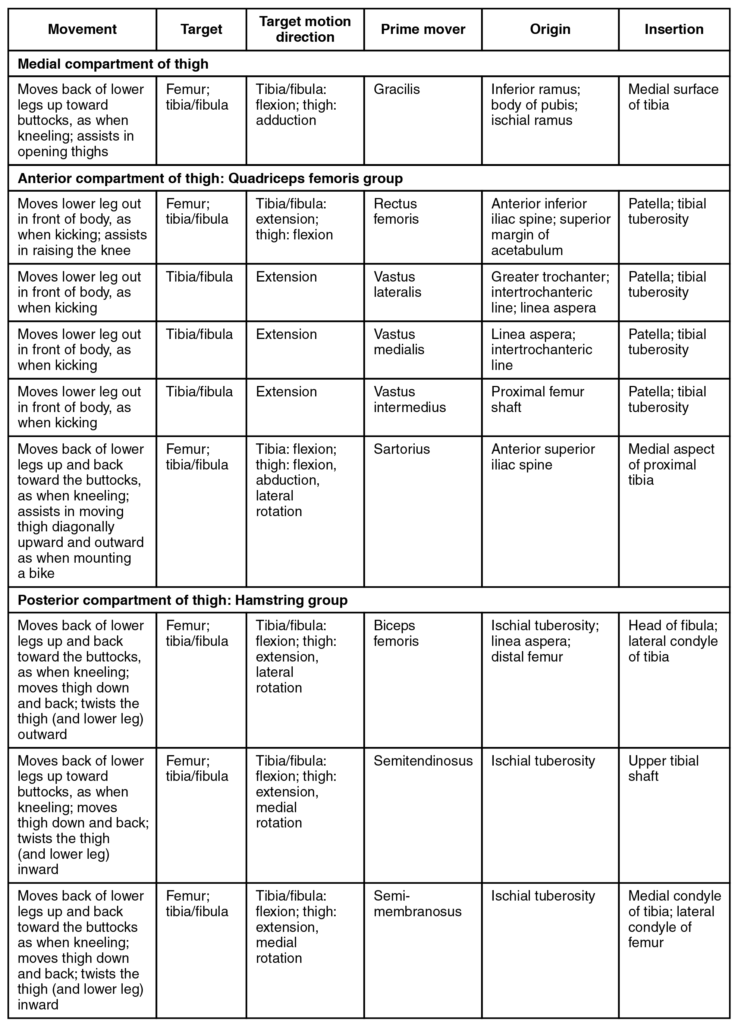
The large and powerful muscles of the hip that move the femur generally originate on the pelvic girdle and insert into the femur. The muscles that move the lower leg typically originate on the femur and insert into the bones of the knee joint. The anterior muscles of the femur extend the lower leg but also aid in flexing the thigh. The posterior muscles of the femur flex the lower leg but also aid in extending the thigh. A combination of gluteal and thigh muscles also adduct, abduct, and rotate the thigh and lower leg.

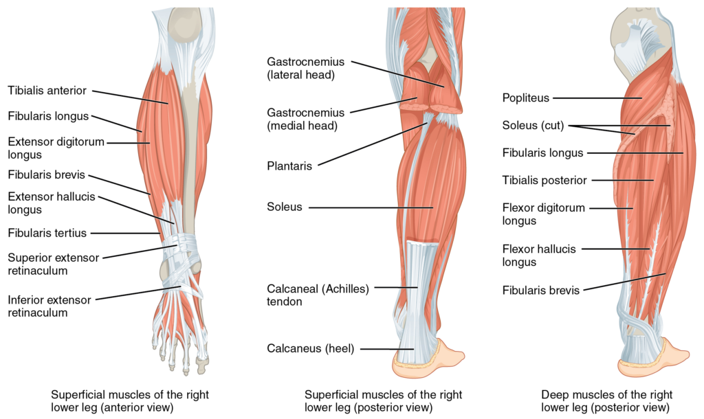
The muscles of the anterior compartment of the lower leg are generally responsible for dorsiflexion, and the muscles of the posterior compartment of the lower leg are generally responsible for plantar flexion. The lateral and medial muscles in both compartments invert, evert, and rotate the foot.

The muscles along the dorsal side of the foot (a) generally extend the toes while the muscles of the plantar side of the foot (b, c, d) generally flex the toes. The plantar muscles exist in four layers, providing the foot the strength to counterbalance the weight of the body. In this diagram, three of the layers are shown from a plantar view beginning with the bottom-most layer just under the plantar skin of the foot (b) and ending with the top-most layer (d) located just inferior to the foot and toe bones.

Nội dung này đang được cập nhật.
Dưới đây là video và các luyện tập (practice) của bài này. Nghe là một kĩ năng khó, đặc biệt là khi chúng ta chưa quen nội dung và chưa có nhạy cảm ngôn ngữ. Nhưng cứ đi thật chậm và đừng bỏ cuộc.
Xem video và cảm nhận nội dung bài. Bạn có thể thả trôi, cảm nhận dòng chảy ngôn ngữ và không nhất thiết phải hiểu toàn bộ bài. Bên dưới là script để bạn khái quát nội dụng và tra từ mới.
Script:
  1. The pelvic girdle attaches the legs to the axial skeleton.
  2. The hip joint is where the pelvic girdle and the leg come together.
  3. The hip is joined to the pelvic girdle by many muscles.
  4. In the gluteal region, the psoas major and iliacus form the iliopsoas.
  5. The large and strong gluteus maximus, gluteus medius, and gluteus minimus extend and abduct the femur.
  6. Along with the gluteus maximus, the tensor fascia lata muscle forms the iliotibial tract.
  7. The lateral rotators of the femur at the hip are the piriformis, obturator internus, obturator externus, superior gemellus, inferior gemellus, and quadratus femoris.
  8. On the medial part of the thigh, the adductor longus, adductor brevis, and adductor magnus adduct the thigh and medially rotate it.
  9. The pectineus muscle adducts and flexes the femur at the hip.
  10. The thigh muscles that move the femur, tibia, and fibula are divided into medial, anterior, and posterior compartments.
  11. The medial compartment includes the adductors, pectineus, and the gracilis.
  12. The anterior compartment comprises the quadriceps femoris, quadriceps tendon, patellar ligament, and the sartorius.
  13. The quadriceps femoris is made of four muscles: the rectus femoris, the vastus lateralis, the vastus medius, and the vastus intermedius, which together extend the knee.
  14. The posterior compartment of the thigh includes the hamstrings: the biceps femoris, semitendinosus, and the semimembranosus, which all flex the knee.
  15. The muscles of the leg that move the foot and toes are divided into anterior, lateral, superficial and deep posterior compartments.
  16. The anterior compartment includes the tibialis anterior, the extensor hallucis longus, the extensor digitorum longus, and the fibularis tertius or peroneus tertius.
  17. The lateral compartment houses the fibularis longus or peroneus longus and the fibularis brevis or peroneus brevis.
Bật video, nghe và điền từ vào chỗ trống.
Dưới đây là phần bàn luận. Bạn có thể tự do đặt câu hỏi, bổ sung kiến thức, và chia sẻ trải nghiệm của mình.
Subscribe
Notify of

0 Comments
Inline Feedbacks
View all comments

Ấn vào ô bên dưới để đánh dấu bạn đã hoàn thành bài học này

Quá dữ! Tiếp tục duy trì phong độ nhé!