Module 8: Anatomy of the Nervous System

Lesson 1: Embryology of the Nervous System

Phôi Thai Học Hệ Thần Kinh

Nội dung bài học:
Mỗi bài học (lesson) bao gồm 4 phần chính: Thuật ngữ, Luyện Đọc, Luyện Nghe, và Bàn Luận.
Sử dụng tính năng:
Bôi hoặc nhấp đôi vào từ, sau đó ấn vào biểu tượng để tra nghĩa từ đó. Khi bạn đưa chuột đến câu (hoặc khi nhấp vào câu trên màn hình điện thoại), gợi ý về cách hiểu câu đó sẽ hiện lên. Cuối cùng, bạn có thể đánh dấu hoàn thành toàn bộ bài học bằng cách ấn vào nút “Hoàn Thành” ở cuối trang.
Đăng ký và đăng nhập
Bạn cần đăng ký và đăng nhập vào tài khoản để lưu quá trình học.
Dưới đây là danh sách những thuật ngữ Y khoa của module Anatomy of the Nervous System.
Khái quát được số lượng thuật ngữ sẽ xuất hiện trong bài đọc và nghe sẽ giúp bạn thoải mái tiêu thụ nội dung hơn. Sau khi hoàn thành nội dung đọc và nghe, bạn hãy quay lại đây và luyện tập (practice) để quen dần các thuật ngữ này. Đừng ép bản thân phải nhớ các thuật ngữ này vội vì bạn sẽ gặp và ôn lại danh sách này trong những bài học (lesson) khác của cùng một module.

Medical Terminology: Anatomy of the Nervous System

abducens nerve
sixth cranial nerve; responsible for contraction of one of the extraocular muscles
alar plate
developmental region of the spinal cord that gives rise to the posterior horn of the gray matter
amygdala
nucleus deep in the temporal lobe of the cerebrum that is related to memory and emotional behavior
anterior column
white matter between the anterior horns of the spinal cord composed of many different groups of axons of both ascending and descending tracts
anterior horn
gray matter of the spinal cord containing multipolar motor neurons, sometimes referred to as the ventral horn
anterior median fissure
deep midline feature of the anterior spinal cord, marking the separation between the right and left sides of the cord
anterior spinal artery
blood vessel from the merged branches of the vertebral arteries that runs along the anterior surface of the spinal cord
arachnoid granulation
outpocket of the arachnoid membrane into the dural sinuses that allows for reabsorption of CSF into the blood
arachnoid mater
middle layer of the meninges named for the spider-web–like trabeculae that extend between it and the pia mater
arachnoid trabeculae
filaments between the arachnoid and pia mater within the subarachnoid space
ascending tract
central nervous system fibers carrying sensory information from the spinal cord or periphery to the brain
axillary nerve
systemic nerve of the arm that arises from the brachial plexus
basal forebrain
nuclei of the cerebrum related to modulation of sensory stimuli and attention through broad projections to the cerebral cortex, loss of which is related to Alzheimer’s disease
basal nuclei
nuclei of the cerebrum (with a few components in the upper brain stem and diencephalon) that are responsible for assessing cortical movement commands and comparing them with the general state of the individual through broad modulatory activity of dopamine neurons; largely related to motor functions, as evidenced through the symptoms of Parkinson’s and Huntington’s diseases
basal plate
developmental region of the spinal cord that gives rise to the lateral and anterior horns of gray matter
basilar artery
blood vessel from the merged vertebral arteries that runs along the dorsal surface of the brain stem
brachial plexus
nerve plexus associated with the lower cervical spinal nerves and first thoracic spinal nerve
brain stem
region of the adult brain that includes the midbrain, pons, and medulla oblongata and develops from the mesencephalon, metencephalon, and myelencephalon of the embryonic brain
Broca’s area
region of the frontal lobe associated with the motor commands necessary for speech production and located only in the cerebral hemisphere responsible for language production, which is the left side in approximately 95 percent of the population
Brodmann’s areas
mapping of regions of the cerebral cortex based on microscopic anatomy that relates specific areas to functional differences, as described by Brodmann in the early 1900s
carotid canal
opening in the temporal bone through which the internal carotid artery enters the cranium
cauda equina
bundle of spinal nerve roots that descend from the lower spinal cord below the first lumbar vertebra and lie within the vertebral cavity; has the appearance of a horse’s tail
caudate
nucleus deep in the cerebrum that is part of the basal nuclei; along with the putamen, it is part of the striatum
central canal
hollow space within the spinal cord that is the remnant of the center of the neural tube
central sulcus
surface landmark of the cerebral cortex that marks the boundary between the frontal and parietal lobes
cephalic flexure
curve in midbrain of the embryo that positions the forebrain ventrally
cerebellum
region of the adult brain connected primarily to the pons that developed from the metencephalon (along with the pons) and is largely responsible for comparing information from the cerebrum with sensory feedback from the periphery through the spinal cord
cerebral aqueduct
connection of the ventricular system between the third and fourth ventricles located in the midbrain
cerebral cortex
outer gray matter covering the forebrain, marked by wrinkles and folds known as gyri and sulci
cerebral hemisphere
one half of the bilaterally symmetrical cerebrum
cerebrum
region of the adult brain that develops from the telencephalon and is responsible for higher neurological functions such as memory, emotion, and consciousness
cervical plexus
nerve plexus associated with the upper cervical spinal nerves
choroid plexus
specialized structures containing ependymal cells lining blood capillaries that filter blood to produce CSF in the four ventricles of the brain
circle of Willis
unique anatomical arrangement of blood vessels around the base of the brain that maintains perfusion of blood into the brain even if one component of the structure is blocked or narrowed
common carotid artery
blood vessel that branches off the aorta (or the brachiocephalic artery on the right) and supplies blood to the head and neck
corpus callosum
large white matter structure that connects the right and left cerebral hemispheres
cranial nerve
one of twelve nerves connected to the brain that are responsible for sensory or motor functions of the head and neck
cranial nerve ganglion
sensory ganglion of cranial nerves
descending tract
central nervous system fibers carrying motor commands from the brain to the spinal cord or periphery
diencephalon
region of the adult brain that retains its name from embryonic development and includes the thalamus and hypothalamus
direct pathway
connections within the basal nuclei from the striatum to the globus pallidus internal segment and substantia nigra pars reticulata that disinhibit the thalamus to increase cortical control of movement
disinhibition
disynaptic connection in which the first synapse inhibits the second cell, which then stops inhibiting the final target
dorsal (posterior) nerve root
axons entering the posterior horn of the spinal cord
dorsal (posterior) root ganglion
sensory ganglion attached to the posterior nerve root of a spinal nerve
dura mater
tough, fibrous, outer layer of the meninges that is attached to the inner surface of the cranium and vertebral column and surrounds the entire CNS
dural sinus
any of the venous structures surrounding the brain, enclosed within the dura mater, which drain blood from the CNS to the common venous return of the jugular veins
endoneurium
innermost layer of connective tissue that surrounds individual axons within a nerve
enteric nervous system
peripheral structures, namely ganglia and nerves, that are incorporated into the digestive system organs
enteric plexus
neuronal plexus in the wall of the intestines, which is part of the enteric nervous system
epineurium
outermost layer of connective tissue that surrounds an entire nerve
epithalamus
region of the diecephalon containing the pineal gland
esophageal plexus
neuronal plexus in the wall of the esophagus that is part of the enteric nervous system
extraocular muscles
six skeletal muscles that control eye movement within the orbit
facial nerve
seventh cranial nerve; responsible for contraction of the facial muscles and for part of the sense of taste, as well as causing saliva production
fascicle
small bundles of nerve or muscle fibers enclosed by connective tissue
femoral nerve
systemic nerve of the anterior leg that arises from the lumbar plexus
fibular nerve
systemic nerve of the posterior leg that begins as part of the sciatic nerve
foramen magnum
large opening in the occipital bone of the skull through which the spinal cord emerges and the vertebral arteries enter the cranium
forebrain
anterior region of the adult brain that develops from the prosencephalon and includes the cerebrum and diencephalon
fourth ventricle
the portion of the ventricular system that is in the region of the brain stem and opens into the subarachnoid space through the median and lateral apertures
frontal eye field
region of the frontal lobe associated with motor commands to orient the eyes toward an object of visual attention
frontal lobe
region of the cerebral cortex directly beneath the frontal bone of the cranium
gastric plexuses
neuronal networks in the wall of the stomach that are part of the enteric nervous system
globus pallidus
nuclei deep in the cerebrum that are part of the basal nuclei and can be divided into the internal and external segments
glossopharyngeal nerve
ninth cranial nerve; responsible for contraction of muscles in the tongue and throat and for part of the sense of taste, as well as causing saliva production
gyrus
ridge formed by convolutions on the surface of the cerebrum or cerebellum
hindbrain
posterior region of the adult brain that develops from the rhombencephalon and includes the pons, medulla oblongata, and cerebellum
hippocampus
gray matter deep in the temporal lobe that is very important for long-term memory formation
hypoglossal nerve
twelfth cranial nerve; responsible for contraction of muscles of the tongue
hypothalamus
major region of the diencephalon that is responsible for coordinating autonomic and endocrine control of homeostasis
indirect pathway
connections within the basal nuclei from the striatum through the globus pallidus external segment and subthalamic nucleus to the globus pallidus internal segment/substantia nigra pars compacta that result in inhibition of the thalamus to decrease cortical control of movement
inferior colliculus
half of the midbrain tectum that is part of the brain stem auditory pathway
inferior olive
nucleus in the medulla that is involved in processing information related to motor control
intercostal nerve
systemic nerve in the thoracic cavity that is found between two ribs
internal carotid artery
branch from the common carotid artery that enters the cranium and supplies blood to the brain
interventricular foramina
openings between the lateral ventricles and third ventricle allowing for the passage of CSF
jugular veins
blood vessels that return “used” blood from the head and neck
kinesthesia
general sensory perception of movement of the body
lateral apertures
pair of openings from the fourth ventricle to the subarachnoid space on either side and between the medulla and cerebellum
lateral column
white matter of the spinal cord between the posterior horn on one side and the axons from the anterior horn on the same side; composed of many different groups of axons, of both ascending and descending tracts, carrying motor commands to and from the brain
lateral horn
region of the spinal cord gray matter in the thoracic, upper lumbar, and sacral regions that is the central component of the sympathetic division of the autonomic nervous system
lateral sulcus
surface landmark of the cerebral cortex that marks the boundary between the temporal lobe and the frontal and parietal lobes
lateral ventricles
portions of the ventricular system that are in the region of the cerebrum
limbic cortex
collection of structures of the cerebral cortex that are involved in emotion, memory, and behavior and are part of the larger limbic system
limbic system
structures at the edge (limit) of the boundary between the forebrain and hindbrain that are most associated with emotional behavior and memory formation
longitudinal fissure
large separation along the midline between the two cerebral hemispheres
lumbar plexus
nerve plexus associated with the lumbar spinal nerves
lumbar puncture
procedure used to withdraw CSF from the lower lumbar region of the vertebral column that avoids the risk of damaging CNS tissue because the spinal cord ends at the upper lumbar vertebrae
median aperture
singular opening from the fourth ventricle into the subarachnoid space at the midline between the medulla and cerebellum
median nerve
systemic nerve of the arm, located between the ulnar and radial nerves
meninges
protective outer coverings of the CNS composed of connective tissue
mesencephalon
primary vesicle of the embryonic brain that does not significantly change through the rest of embryonic development and becomes the midbrain
metencephalon
secondary vesicle of the embryonic brain that develops into the pons and the cerebellum
midbrain
middle region of the adult brain that develops from the mesencephalon
myelencephalon
secondary vesicle of the embryonic brain that develops into the medulla
nerve plexus
network of nerves without neuronal cell bodies included
neural crest
tissue that detaches from the edges of the neural groove and migrates through the embryo to develop into peripheral structures of both nervous and non-nervous tissues
neural fold
elevated edge of the neural groove
neural groove
region of the neural plate that folds into the dorsal surface of the embryo and closes off to become the neural tube
neural plate
thickened layer of neuroepithelium that runs longitudinally along the dorsal surface of an embryo and gives rise to nervous system tissue
neural tube
precursor to structures of the central nervous system, formed by the invagination and separation of neuroepithelium
neuraxis
central axis to the nervous system, from the posterior to anterior ends of the neural tube; the inferior tip of the spinal cord to the anterior surface of the cerebrum
occipital lobe
region of the cerebral cortex directly beneath the occipital bone of the cranium
occipital sinuses
dural sinuses along the edge of the occipital lobes of the cerebrum
oculomotor nerve
third cranial nerve; responsible for contraction of four of the extraocular muscles, the muscle in the upper eyelid, and pupillary constriction
olfaction
special sense responsible for smell, which has a unique, direct connection to the cerebrum
olfactory nerve
first cranial nerve; responsible for the sense of smell
optic nerve
second cranial nerve; responsible for visual sensation
orthostatic reflex
sympathetic function that maintains blood pressure when standing to offset the increased effect of gravity
paravertebral ganglia
autonomic ganglia superior to the sympathetic chain ganglia
parietal lobe
region of the cerebral cortex directly beneath the parietal bone of the cranium
parieto-occipital sulcus
groove in the cerebral cortex representing the border between the parietal and occipital cortices
perineurium
layer of connective tissue surrounding fascicles within a nerve
phrenic nerve
systemic nerve from the cervical plexus that innervates the diaphragm
pia mater
thin, innermost membrane of the meninges that directly covers the surface of the CNS
plexus
network of nerves or nervous tissue
postcentral gyrus
primary motor cortex located in the frontal lobe of the cerebral cortex
posterior columns
white matter of the spinal cord that lies between the posterior horns of the gray matter, sometimes referred to as the dorsal column; composed of axons of ascending tracts that carry sensory information up to the brain
posterior horn
gray matter region of the spinal cord in which sensory input arrives, sometimes referred to as the dorsal horn
posterior median sulcus
midline feature of the posterior spinal cord, marking the separation between right and left sides of the cord
posterolateral sulcus
feature of the posterior spinal cord marking the entry of posterior nerve roots and the separation between the posterior and lateral columns of the white matter
precentral gyrus
ridge just posterior to the central sulcus, in the parietal lobe, where somatosensory processing initially takes place in the cerebrum
prefrontal lobe
specific region of the frontal lobe anterior to the more specific motor function areas, which can be related to the early planning of movements and intentions to the point of being personality-type functions
premotor area
region of the frontal lobe responsible for planning movements that will be executed through the primary motor cortex
prevertebral ganglia
autonomic ganglia that are anterior to the vertebral column and functionally related to the sympathetic chain ganglia
primary vesicle
initial enlargements of the anterior neural tube during embryonic development that develop into the forebrain, midbrain, and hindbrain
proprioception
general sensory perceptions providing information about location and movement of body parts; the “sense of the self”
prosencephalon
primary vesicle of the embryonic brain that develops into the forebrain, which includes the cerebrum and diencephalon
putamen
nucleus deep in the cerebrum that is part of the basal nuclei; along with the caudate, it is part of the striatum
radial nerve
systemic nerve of the arm, the distal component of which is located near the radial bone
reticular formation
diffuse region of gray matter throughout the brain stem that regulates sleep, wakefulness, and states of consciousness
rhombencephalon
primary vesicle of the embryonic brain that develops into the hindbrain, which includes the pons, cerebellum, and medulla
sacral plexus
nerve plexus associated with the lower lumbar and sacral spinal nerves
saphenous nerve
systemic nerve of the lower anterior leg that is a branch from the femoral nerve
sciatic nerve
systemic nerve from the sacral plexus that is a combination of the tibial and fibular nerves and extends across the hip joint and gluteal region into the upper posterior leg
sciatica
painful condition resulting from inflammation or compression of the sciatic nerve or any of the spinal nerves that contribute to it
secondary vesicle
five vesicles that develop from primary vesicles, continuing the process of differentiation of the embryonic brain
sigmoid sinuses
dural sinuses that drain directly into the jugular veins
somatosensation
general senses related to the body, usually thought of as the senses of touch, which would include pain, temperature, and proprioception
spinal accessory nerve
eleventh cranial nerve; responsible for contraction of neck muscles
spinal nerve
one of 31 nerves connected to the spinal cord
straight sinus
dural sinus that drains blood from the deep center of the brain to collect with the other sinuses
striatum
the caudate and putamen collectively, as part of the basal nuclei, which receive input from the cerebral cortex
subarachnoid space
space between the arachnoid mater and pia mater that contains CSF and the fibrous connections of the arachnoid trabeculae
subcortical nucleus
all the nuclei beneath the cerebral cortex, including the basal nuclei and the basal forebrain
substantia nigra pars compacta
nuclei within the basal nuclei that release dopamine to modulate the function of the striatum; part of the motor pathway
substantia nigra pars reticulata
nuclei within the basal nuclei that serve as an output center of the nuclei; part of the motor pathway
subthalamus
nucleus within the basal nuclei that is part of the indirect pathway
sulcus
groove formed by convolutions in the surface of the cerebral cortex
superior colliculus
half of the midbrain tectum that is responsible for aligning visual, auditory, and somatosensory spatial perceptions
superior sagittal sinus
dural sinus that runs along the top of the longitudinal fissure and drains blood from the majority of the outer cerebrum
sympathetic chain ganglia
autonomic ganglia in a chain along the anterolateral aspect of the vertebral column that are responsible for contributing to homeostatic mechanisms of the autonomic nervous system
systemic nerve
nerve in the periphery distal to a nerve plexus or spinal nerve
tectum
region of the midbrain, thought of as the roof of the cerebral aqueduct, which is subdivided into the inferior and superior colliculi
tegmentum
region of the midbrain, thought of as the floor of the cerebral aqueduct, which continues into the pons and medulla as the floor of the fourth ventricle
telencephalon
secondary vesicle of the embryonic brain that develops into the cerebrum
temporal lobe
region of the cerebral cortex directly beneath the temporal bone of the cranium
terminal ganglion
autonomic ganglia that are near or within the walls of organs that are responsible for contributing to homeostatic mechanisms of the autonomic nervous system
thalamus
major region of the diencephalon that is responsible for relaying information between the cerebrum and the hindbrain, spinal cord, and periphery
third ventricle
portion of the ventricular system that is in the region of the diencephalon
tibial nerve
systemic nerve of the posterior leg that begins as part of the sciatic nerve
transverse sinuses
dural sinuses that drain along either side of the occipital–cerebellar space
trigeminal ganglion
sensory ganglion that contributes sensory fibers to the trigeminal nerve
trigeminal nerve
fifth cranial nerve; responsible for cutaneous sensation of the face and contraction of the muscles of mastication
trochlear nerve
fourth cranial nerve; responsible for contraction of one of the extraocular muscles
ulnar nerve
systemic nerve of the arm located close to the ulna, a bone of the forearm
vagus nerve
tenth cranial nerve; responsible for the autonomic control of organs in the thoracic and upper abdominal cavities
ventral (anterior) nerve root
axons emerging from the anterior or lateral horns of the spinal cord
ventricles
remnants of the hollow center of the neural tube that are spaces for cerebrospinal fluid to circulate through the brain
vertebral arteries
arteries that ascend along either side of the vertebral column through the transverse foramina of the cervical vertebrae and enter the cranium through the foramen magnum
vestibulocochlear nerve
eighth cranial nerve; responsible for the sensations of hearing and balance
Nội dung này đang được cập nhật.
Dưới đây là các bài văn nằm ở bên trái. Ở bên phải là các bài luyện tập (practice) để đánh giá khả năng đọc hiểu của bạn. Sẽ khó khăn trong thời gian đầu nếu vốn từ vựng của bạn còn hạn chế, đặc biệt là từ vựng Y khoa. Hãy kiên nhẫn và đọc nhiều nhất có kể, lượng kiến thức tích tụ dần sẽ giúp bạn đọc thoải mái hơn.
The brain is a complex organ composed of gray parts and white matter, which can be hard to distinguish. Starting from an embryologic perspective allows you to understand more easily how the parts relate to each other. The embryonic nervous system begins as a very simple structure—essentially just a straight line, which then gets increasingly complex. Looking at the development of the nervous system with a couple of early snapshots makes it easier to understand the whole complex system.

Many structures that appear to be adjacent in the adult brain are not connected, and the connections that exist may seem arbitrary. But there is an underlying order to the system that comes from how different parts develop. By following the developmental pattern, it is possible to learn what the major regions of the nervous system are.
To begin, a sperm cell and an egg cell fuse to become a fertilized egg. The fertilized egg cell, or zygote, starts dividing to generate the cells that make up an entire organism. Sixteen days after fertilization, the developing embryo’s cells belong to one of three germ layers that give rise to the different tissues in the body. The endoderm, or inner tissue, is responsible for generating the lining tissues of various spaces within the body, such as the mucosae of the digestive and respiratory systems. The mesoderm, or middle tissue, gives rise to most of the muscle and connective tissues. Finally the ectoderm, or outer tissue, develops into the integumentary system (the skin) and the nervous system. It is probably not difficult to see that the outer tissue of the embryo becomes the outer covering of the body. But how is it responsible for the nervous system?

As the embryo develops, a portion of the ectoderm differentiates into a specialized region of neuroectoderm, which is the precursor for the tissue of the nervous system. Molecular signals induce cells in this region to differentiate into the neuroepithelium, forming a neural plate. The cells then begin to change shape, causing the tissue to buckle and fold inward (Figure 1). A neural groove forms, visible as a line along the dorsal surface of the embryo. The ridge-like edge on either side of the neural groove is referred as the neural fold. As the neural folds come together and converge, the underlying structure forms into a tube just beneath the ectoderm called the neural tube. Cells from the neural folds then separate from the ectoderm to form a cluster of cells referred to as the neural crest, which runs lateral to the neural tube. The neural crest migrates away from the nascent, or embryonic, central nervous system (CNS) that will form along the neural groove and develops into several parts of the peripheral nervous system (PNS), including the enteric nervous tissue. Many tissues that are not part of the nervous system also arise from the neural crest, such as craniofacial cartilage and bone, and melanocytes.

At this point, the early nervous system is a simple, hollow tube. It runs from the anterior end of the embryo to the posterior end. Beginning at 25 days, the anterior end develops into the brain, and the posterior portion becomes the spinal cord. This is the most basic arrangement of tissue in the nervous system, and it gives rise to the more complex structures by the fourth week of development.
As the anterior end of the neural tube starts to develop into the brain, it undergoes a couple of enlargements; the result is the production of sac-like vesicles. Similar to a child’s balloon animal, the long, straight neural tube begins to take on a new shape. Three vesicles form at the first stage, which are called primary vesicles. These vesicles are given names that are based on Greek words, the main root word being enkephalon, which means “brain” (en- = “inside”; kephalon = “head”). The prefix to each generally corresponds to its position along the length of the developing nervous system.

The prosencephalon (pros- = “in front”) is the forward-most vesicle, and the term can be loosely translated to mean forebrain. The mesencephalon (mes- = “middle”) is the next vesicle, which can be called the midbrain. The third vesicle at this stage is the rhombencephalon. The first part of this word is also the root of the word rhombus, which is a geometrical figure with four sides of equal length (a square is a rhombus with 90° angles). Whereas prosencephalon and mesencephalon translate into the English words forebrain and midbrain, there is not a word for “four-sided-figure-brain.” However, the third vesicle can be called the hindbrain. One way of thinking about how the brain is arranged is to use these three regions—forebrain, midbrain, and hindbrain—which are based on the primary vesicle stage of development (Figure 2a).
The brain continues to develop, and the vesicles differentiate further (see Figure 2b). The three primary vesicles become five secondary vesicles. The prosencephalon enlarges into two new vesicles called the telencephalon and the diencephalon. The telecephalon will become the cerebrum. The diencephalon gives rise to several adult structures; two that will be important are the thalamus and the hypothalamus. In the embryonic diencephalon, a structure known as the eye cup develops, which will eventually become the retina, the nervous tissue of the eye called the retina. This is a rare example of nervous tissue developing as part of the CNS structures in the embryo, but becoming a peripheral structure in the fully formed nervous system.

The mesencephalon does not differentiate into any finer divisions. The midbrain is an established region of the brain at the primary vesicle stage of development and remains that way. The rest of the brain develops around it and constitutes a large percentage of the mass of the brain. Dividing the brain into forebrain, midbrain, and hindbrain is useful in considering its developmental pattern, but the midbrain is a small proportion of the entire brain, relatively speaking.

The rhombencephalon develops into the metencephalon and myelencephalon. The metencephalon corresponds to the adult structure known as the pons and also gives rise to the cerebellum. The cerebellum (from the Latin meaning “little brain”) accounts for about 10 percent of the mass of the brain and is an important structure in itself. The most significant connection between the cerebellum and the rest of the brain is at the pons, because the pons and cerebellum develop out of the same vesicle. The myelencephalon corresponds to the adult structure known as the medulla oblongata. The structures that come from the mesencephalon and rhombencephalon, except for the cerebellum, are collectively considered the brain stem, which specifically includes the midbrain, pons, and medulla.
While the brain is developing from the anterior neural tube, the spinal cord is developing from the posterior neural tube. However, its structure does not differ from the basic layout of the neural tube. It is a long, straight cord with a small, hollow space down the center. The neural tube is defined in terms of its anterior versus posterior portions, but it also has a dorsal–ventral dimension. As the neural tube separates from the rest of the ectoderm, the side closest to the surface is dorsal, and the deeper side is ventral.

As the spinal cord develops, the cells making up the wall of the neural tube proliferate and differentiate into the neurons and glia of the spinal cord. The dorsal tissues will be associated with sensory functions, and the ventral tissues will be associated with motor functions.
Embryonic development can help in understanding the structure of the adult brain because it establishes a framework on which more complex structures can be built. First, the neural tube establishes the anterior–posterior dimension of the nervous system, which is called the neuraxis. The embryonic nervous system in mammals can be said to have a standard arrangement. Humans (and other primates, to some degree) make this complicated by standing up and walking on two legs. The anterior–posterior dimension of the neuraxis overlays the superior–inferior dimension of the body. However, there is a major curve between the brain stem and forebrain, which is called the cephalic flexure. Because of this, the neuraxis starts in an inferior position—the end of the spinal cord—and ends in an anterior position, the front of the cerebrum. If this is confusing, just imagine a four-legged animal standing up on two legs. Without the flexure in the brain stem, and at the top of the neck, that animal would be looking straight up instead of straight in front (Figure 3).

In summary, the primary vesicles help to establish the basic regions of the nervous system: forebrain, midbrain, and hindbrain. These divisions are useful in certain situations, but they are not equivalent regions. The midbrain is small compared with the hindbrain and particularly the forebrain. The secondary vesicles go on to establish the major regions of the adult nervous system that will be followed in this text. The telencephalon is the cerebrum, which is the major portion of the human brain. The diencephalon continues to be referred to by this Greek name, because there is no better term for it (dia- = “through”). The diencephalon is between the cerebrum and the rest of the nervous system and can be described as the region through which all projections have to pass between the cerebrum and everything else. The brain stem includes the midbrain, pons, and medulla, which correspond to the mesencephalon, metencephalon, and myelencephalon. The cerebellum, being a large portion of the brain, is considered a separate region. Table 1 connects the different stages of development to the adult structures of the CNS.

One other benefit of considering embryonic development is that certain connections are more obvious because of how these adult structures are related. The retina, which began as part of the diencephalon, is primarily connected to the diencephalon. The eyes are just inferior to the anterior-most part of the cerebrum, but the optic nerve extends back to the thalamus as the optic tract, with branches into a region of the hypothalamus. There is also a connection of the optic tract to the midbrain, but the mesencephalon is adjacent to the diencephalon, so that is not difficult to imagine. The cerebellum originates out of the metencephalon, and its largest white matter connection is to the pons, also from the metencephalon. There are connections between the cerebellum and both the medulla and midbrain, which are adjacent structures in the secondary vesicle stage of development. In the adult brain, the cerebellum seems close to the cerebrum, but there is no direct connection between them.

Another aspect of the adult CNS structures that relates to embryonic development is the ventricles—open spaces within the CNS where cerebrospinal fluid circulates. They are the remnant of the hollow center of the neural tube. The four ventricles and the tubular spaces associated with them can be linked back to the hollow center of the embryonic brain (see Table 1).

OpenStax. (2022). Anatomy and Physiology 2e. Rice University. Retrieved June 15, 2023. ISBN-13: 978-1-711494-06-7 (Hardcover) ISBN-13: 978-1-711494-05-0 (Paperback) ISBN-13: 978-1-951693-42-8 (Digital). License: Attribution 4.0 International (CC BY 4.0). Access for free at openstax.org.

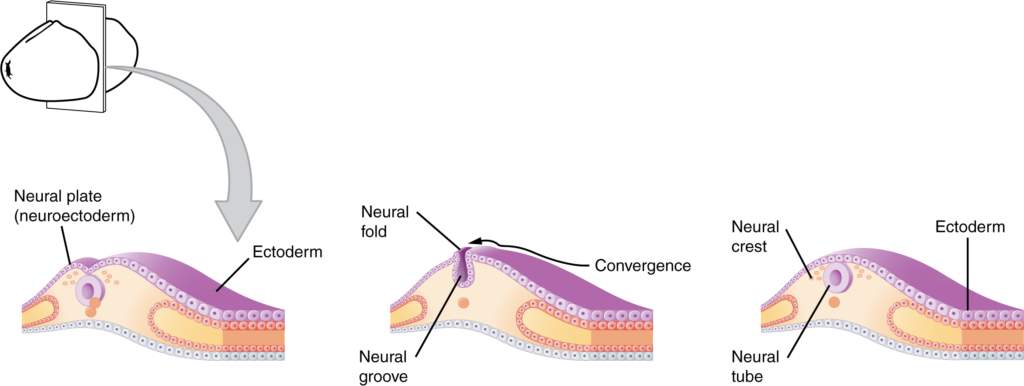
The neuroectoderm begins to fold inward to form the neural groove. As the two sides of the neural groove converge, they form the neural tube, which lies beneath the ectoderm. The anterior end of the neural tube will develop into the brain, and the posterior portion will become the spinal cord. The neural crest develops into peripheral structures.

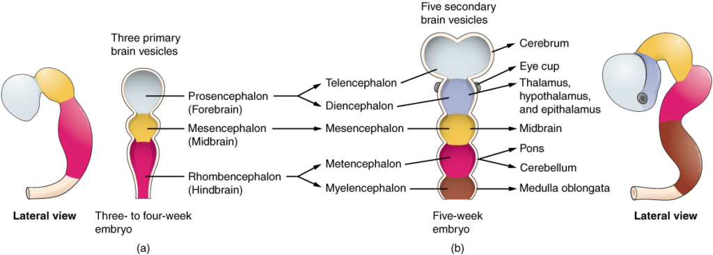
The embryonic brain develops complexity through enlargements of the neural tube called vesicles; (a) The primary vesicle stage has three regions, and (b) the secondary vesicle stage has five regions.

The mammalian nervous system is arranged with the neural tube running along an anterior to posterior axis, from nose to tail for a four-legged animal like a dog. Humans, as two-legged animals, have a bend in the neuraxis between the brain stem and the diencephalon, along with a bend in the neck, so that the eyes and the face are oriented forward.

Neural tubePrimary vesicle stageSecondary vesicle stageAdult structuresVentricles
Anterior neural tubeProsencephalonTelencephalonCerebrumLateral ventricles
Anterior neural tubeProsencephalonDiencephalonDiencephalonThird ventricle
Anterior neural tubeMesencephalonMesencephalonMidbrainCerebral aqueduct
Anterior neural tubeRhombencephalonMetencephalonPons cerebellumFourth ventricle
Anterior neural tubeRhombencephalonMyelencephalonMedullaFourth ventricle
Posterior neural tubeSpinal cordCentral canal
Nội dung này đang được cập nhật.
Dưới đây là video và các luyện tập (practice) của bài này. Nghe là một kĩ năng khó, đặc biệt là khi chúng ta chưa quen nội dung và chưa có nhạy cảm ngôn ngữ. Nhưng cứ đi thật chậm và đừng bỏ cuộc.
Xem video và cảm nhận nội dung bài. Bạn có thể thả trôi, cảm nhận dòng chảy ngôn ngữ và không nhất thiết phải hiểu toàn bộ bài. Bên dưới là script để bạn khái quát nội dụng và tra từ mới.
Script:
  1. The development of the nervous system starts early in embryonic development.
  2. The outer layer of the embryo, the ectoderm, gives rise to the skin and the nervous system.
  3. A specialized region of this layer, the neuroectoderm, becomes a groove that folds in and becomes the neural tube beneath the dorsal surface of the embryo.
  4. The anterior end of the neural tube develops into the brain, and the posterior region becomes the spinal cord.
  5. When the neural groove closes off, tissues at its edges are known as the neural crest.
  6. The neural crest migrates through the embryo, giving rise to peripheral nervous system structures and some non-nervous tissues.
  7. The brain develops from this early tube structure and gives rise to specific regions of the adult brain.
  8. As the neural tube grows and differentiates, it enlarges into three vesicles that correspond to the forebrain, midbrain, and hindbrain regions of the adult brain.
  9. Later in development, two of these three vesicles differentiate further, resulting in five vesicles.
  10. Those five vesicles can be aligned with the four major regions of the adult brain.
  11. The cerebrum is formed directly from the telencephalon.
  12. The diencephalon is the only region that keeps its embryonic name.
  13. The mesencephalon, metencephalon, and myelencephalon become the brain stem.
  14. The cerebellum also develops from the metencephalon and is a separate region of the adult brain.
  15. The spinal cord develops out of the rest of the neural tube and retains the tube structure, with the nervous tissue thickening and the hollow center becoming a very small central canal through the cord.
  16. The rest of the hollow center of the neural tube corresponds to open spaces within the brain called the ventricles, where cerebrospinal fluid is found.
Bật video, nghe và điền từ vào chỗ trống.
Dưới đây là phần bàn luận. Bạn có thể tự do đặt câu hỏi, bổ sung kiến thức, và chia sẻ trải nghiệm của mình.
Subscribe
Notify of

0 Comments
Inline Feedbacks
View all comments

Ấn vào ô bên dưới để đánh dấu bạn đã hoàn thành bài học này

Quá dữ! Tiếp tục duy trì phong độ nhé!