Module 7: The Nervous System and Nervous Tissue

Lesson 5: Communication Between Neurons

Giao Tiếp Giữa Các Tế Bào Thần Kinh

Nội dung bài học:
Mỗi bài học (lesson) bao gồm 4 phần chính: Thuật ngữ, Luyện Đọc, Luyện Nghe, và Bàn Luận.
Sử dụng tính năng:
Bôi hoặc nhấp đôi vào từ, sau đó ấn vào biểu tượng để tra nghĩa từ đó. Khi bạn đưa chuột đến câu (hoặc khi nhấp vào câu trên màn hình điện thoại), gợi ý về cách hiểu câu đó sẽ hiện lên. Cuối cùng, bạn có thể đánh dấu hoàn thành toàn bộ bài học bằng cách ấn vào nút “Hoàn Thành” ở cuối trang.
Đăng ký và đăng nhập
Bạn cần đăng ký và đăng nhập vào tài khoản để lưu quá trình học.
Dưới đây là danh sách những thuật ngữ Y khoa của module The Nervous System and Nervous Tissue.
Khái quát được số lượng thuật ngữ sẽ xuất hiện trong bài đọc và nghe sẽ giúp bạn thoải mái tiêu thụ nội dung hơn. Sau khi hoàn thành nội dung đọc và nghe, bạn hãy quay lại đây và luyện tập (practice) để quen dần các thuật ngữ này. Đừng ép bản thân phải nhớ các thuật ngữ này vội vì bạn sẽ gặp và ôn lại danh sách này trong những bài học (lesson) khác của cùng một module.

Medical Terminology: The Nervous System and Nervous Tissue

absolute refractory period
time during an action period when another action potential cannot be generated because the voltage-gated Na+ channel is inactivated
action potential
change in voltage of a cell membrane in response to a stimulus that results in transmission of an electrical signal; unique to neurons and muscle fibers
activation gate
part of the voltage-gated Na+ channel that opens when the membrane voltage reaches threshold
astrocyte
glial cell type of the CNS that provides support for neurons and maintains the blood-brain barrier
autonomic nervous system (ANS)
functional division of the nervous system that is responsible for homeostatic reflexes that coordinate control of cardiac and smooth muscle, as well as glandular tissue
axon
single process of the neuron that carries an electrical signal (action potential) away from the cell body toward a target cell
axon hillock
tapering of the neuron cell body that gives rise to the axon
axon segment
single stretch of the axon insulated by myelin and bounded by nodes of Ranvier at either end (except for the first, which is after the initial segment, and the last, which is followed by the axon terminal)
axon terminal
end of the axon, where there are usually several branches extending toward the target cell
axoplasm
cytoplasm of an axon, which is different in composition than the cytoplasm of the neuronal cell body
biogenic amine
class of neurotransmitters that are enzymatically derived from amino acids but no longer contain a carboxyl group
bipolar
shape of a neuron with two processes extending from the neuron cell body—the axon and one dendrite
blood-brain barrier (BBB)
physiological barrier between the circulatory system and the central nervous system that establishes a privileged blood supply, restricting the flow of substances into the CNS
brain
the large organ of the central nervous system composed of white and gray matter, contained within the cranium and continuous with the spinal cord
central nervous system (CNS)
anatomical division of the nervous system located within the cranial and vertebral cavities, namely the brain and spinal cord
cerebral cortex
outermost layer of gray matter in the brain, where conscious perception takes place
cerebrospinal fluid (CSF)
circulatory medium within the CNS that is produced by ependymal cells in the choroid plexus filtering the blood
chemical synapse
connection between two neurons, or between a neuron and its target, where a neurotransmitter diffuses across a very short distance
cholinergic system
neurotransmitter system of acetylcholine, which includes its receptors and the enzyme acetylcholinesterase
choroid plexus
specialized structure containing ependymal cells that line blood capillaries and filter blood to produce CSF in the four ventricles of the brain
continuous conduction
slow propagation of an action potential along an unmyelinated axon owing to voltage-gated Na+ channels located along the entire length of the cell membrane
dendrite
one of many branchlike processes that extends from the neuron cell body and functions as a contact for incoming signals (synapses) from other neurons or sensory cells
depolarization
change in a cell membrane potential from rest toward zero
effector protein
enzyme that catalyzes the generation of a new molecule, which acts as the intracellular mediator of the signal that binds to the receptor
electrical synapse
connection between two neurons, or any two electrically active cells, where ions flow directly through channels spanning their adjacent cell membranes
electrochemical exclusion
principle of selectively allowing ions through a channel on the basis of their charge
enteric nervous system (ENS)
neural tissue associated with the digestive system that is responsible for nervous control through autonomic connections
ependymal cell
glial cell type in the CNS responsible for producing cerebrospinal fluid
excitable membrane
cell membrane that regulates the movement of ions so that an electrical signal can be generated
excitatory postsynaptic potential (EPSP)
graded potential in the postsynaptic membrane that is the result of depolarization and makes an action potential more likely to occur
G protein
guanosine triphosphate (GTP) hydrolase that physically moves from the receptor protein to the effector protein to activate the latter
ganglion
localized collection of neuron cell bodies in the peripheral nervous system
gated
property of a channel that determines how it opens under specific conditions, such as voltage change or physical deformation
generator potential
graded potential from dendrites of a unipolar cell which generates the action potential in the initial segment of that cell’s axon
glial cell
one of the various types of neural tissue cells responsible for maintenance of the tissue, and largely responsible for supporting neurons
graded potential
change in the membrane potential that varies in size, depending on the size of the stimulus that elicits it
gray matter
regions of the nervous system containing cell bodies of neurons with few or no myelinated axons; actually may be more pink or tan in color, but called gray in contrast to white matter
inactivation gate
part of a voltage-gated Na+ channel that closes when the membrane potential reaches +30 mV
inhibitory postsynaptic potential (IPSP)
graded potential in the postsynaptic membrane that is the result of hyperpolarization and makes an action potential less likely to occur
initial segment
first part of the axon as it emerges from the axon hillock, where the electrical signals known as action potentials are generated
integration
nervous system function that combines sensory perceptions and higher cognitive functions (memories, learning, emotion, etc.) to produce a response
ionotropic receptor
neurotransmitter receptor that acts as an ion channel gate, and opens by the binding of the neurotransmitter
leakage channel
ion channel that opens randomly and is not gated to a specific event, also known as a non-gated channel
ligand-gated channels
another name for an ionotropic receptor for which a neurotransmitter is the ligand
lower motor neuron
second neuron in the motor command pathway that is directly connected to the skeletal muscle
mechanically gated channel
ion channel that opens when a physical event directly affects the structure of the protein
membrane potential
distribution of charge across the cell membrane, based on the charges of ions
metabotropic receptor
neurotransmitter receptor that involves a complex of proteins that cause metabolic changes in a cell
microglia
glial cell type in the CNS that serves as the resident component of the immune system
multipolar
shape of a neuron that has multiple processes—the axon and two or more dendrites
muscarinic receptor
type of acetylcholine receptor protein that is characterized by also binding to muscarine and is a metabotropic receptor
myelin
lipid-rich insulating substance surrounding the axons of many neurons, allowing for faster transmission of electrical signals
myelin sheath
lipid-rich layer of insulation that surrounds an axon, formed by oligodendrocytes in the CNS and Schwann cells in the PNS; facilitates the transmission of electrical signals
nerve
cord-like bundle of axons located in the peripheral nervous system that transmits sensory input and response output to and from the central nervous system
neuron
neural tissue cell that is primarily responsible for generating and propagating electrical signals into, within, and out of the nervous system
neuropeptide
neurotransmitter type that includes protein molecules and shorter chains of amino acids
neurotransmitter
chemical signal that is released from the synaptic end bulb of a neuron to cause a change in the target cell
nicotinic receptor
type of acetylcholine receptor protein that is characterized by also binding to nicotine and is an ionotropic receptor
node of Ranvier
gap between two myelinated regions of an axon, allowing for strengthening of the electrical signal as it propagates down the axon
nonspecific channel
channel that is not specific to one ion over another, such as a nonspecific cation channel that allows any positively charged ion across the membrane
nucleus
in the nervous system, a localized collection of neuron cell bodies that are functionally related; a “center” of neural function
oligodendrocyte
glial cell type in the CNS that provides the myelin insulation for axons in tracts
peripheral nervous system (PNS)
anatomical division of the nervous system that is largely outside the cranial and vertebral cavities, namely all parts except the brain and spinal cord
postsynaptic potential (PSP)
graded potential in the postsynaptic membrane caused by the binding of neurotransmitter to protein receptors
precentral gyrus of the frontal cortex
region of the cerebral cortex responsible for generating motor commands, where the upper motor neuron cell body is located
process
in cells, an extension of a cell body; in the case of neurons, this includes the axon and dendrites
propagation
movement of an action potential along the length of an axon
receptor potential
graded potential in a specialized sensory cell that directly causes the release of neurotransmitter without an intervening action potential
refractory period
time after the initiation of an action potential when another action potential cannot be generated
relative refractory period
time during the refractory period when a new action potential can only be initiated by a stronger stimulus than the current action potential because voltage-gated K+ channels are not closed
repolarization
return of the membrane potential to its normally negative voltage at the end of the action potential
resistance
property of an axon that relates to the ability of particles to diffuse through the cytoplasm; this is inversely proportional to the fiber diameter
response
nervous system function that causes a target tissue (muscle or gland) to produce an event as a consequence to stimuli
resting membrane potential
the difference in voltage measured across a cell membrane under steady-state conditions, typically -70 mV
saltatory conduction
quick propagation of the action potential along a myelinated axon owing to voltage-gated Na+ channels being present only at the nodes of Ranvier
satellite cell
glial cell type in the PNS that provides support for neurons in the ganglia
Schwann cell
glial cell type in the PNS that provides the myelin insulation for axons in nerves
sensation
nervous system function that receives information from the environment and translates it into the electrical signals of nervous tissue
size exclusion
principle of selectively allowing ions through a channel on the basis of their relative size
soma
in neurons, that portion of the cell that contains the nucleus; the cell body, as opposed to the cell processes (axons and dendrites)
somatic nervous system (SNS)
functional division of the nervous system that is concerned with conscious perception, voluntary movement, and skeletal muscle reflexes
spatial summation
combination of graded potentials across the neuronal cell membrane caused by signals from separate presynaptic elements that add up to initiate an action potential
spinal cord
organ of the central nervous system found within the vertebral cavity and connected with the periphery through spinal nerves; mediates reflex behaviors
stimulus
an event in the external or internal environment that registers as activity in a sensory neuron
summate
to add together, as in the cumulative change in postsynaptic potentials toward reaching threshold in the membrane, either across a span of the membrane or over a certain amount of time
synapse
narrow junction across which a chemical signal passes from neuron to the next, initiating a new electrical signal in the target cell
synaptic cleft
small gap between cells in a chemical synapse where neurotransmitter diffuses from the presynaptic element to the postsynaptic element
synaptic end bulb
swelling at the end of an axon where neurotransmitter molecules are released onto a target cell across a synapse
temporal summation
combination of graded potentials at the same location on a neuron resulting in a strong signal from one input
thalamus
region of the central nervous system that acts as a relay for sensory pathways
thermoreceptor
type of sensory receptor capable of transducing temperature stimuli into neural action potentials
threshold
membrane voltage at which an action potential is initiated
tract
bundle of axons in the central nervous system having the same function and point of origin
unipolar
shape of a neuron which has only one process that includes both the axon and dendrite
upper motor neuron
first neuron in the motor command pathway with its cell body in the cerebral cortex that synapses on the lower motor neuron in the spinal cord
ventricle
central cavity within the brain where CSF is produced and circulates
voltage-gated channel
ion channel that opens because of a change in the charge distributed across the membrane where it is located
white matter
regions of the nervous system containing mostly myelinated axons, making the tissue appear white because of the high lipid content of myelin
Nội dung này đang được cập nhật.
Dưới đây là các bài văn nằm ở bên trái. Ở bên phải là các bài luyện tập (practice) để đánh giá khả năng đọc hiểu của bạn. Sẽ khó khăn trong thời gian đầu nếu vốn từ vựng của bạn còn hạn chế, đặc biệt là từ vựng Y khoa. Hãy kiên nhẫn và đọc nhiều nhất có kể, lượng kiến thức tích tụ dần sẽ giúp bạn đọc thoải mái hơn.
The electrical changes taking place within a neuron, as described in the previous section, are similar to a light switch being turned on. A stimulus starts the depolarization, but the action potential runs on its own once a threshold has been reached. The question is now, “What flips the light switch on?” Temporary changes to the cell membrane voltage can result from neurons receiving information from the environment, or from the action of one neuron on another. These special types of potentials influence a neuron and determine whether an action potential will occur or not. Many of these transient signals originate at the synapse.
Local changes in the membrane potential are called graded potentials and are usually associated with the dendrites of a neuron. The amount of change in the membrane potential is determined by the size of the stimulus that causes it. In the example of testing the temperature of the shower, slightly warm water would only initiate a small change in a thermoreceptor, whereas hot water would cause a large amount of change in the membrane potential.

Graded potentials can be of two sorts, either they are depolarizing or hyperpolarizing (Figure 12.25). For a membrane at the resting potential, a graded potential represents a change in that voltage either above -70 mV or below -70 mV. Depolarizing graded potentials are often the result of Na+ or Ca2+ entering the cell. Both of these ions have higher concentrations outside the cell than inside; because they have a positive charge, they will move into the cell causing it to become less negative relative to the outside. Hyperpolarizing graded potentials can be caused by K+ leaving the cell or Cl- entering the cell. If a positive charge moves out of a cell, the cell becomes more negative; if a negative charge enters the cell, the same thing happens.

A. Types of Graded Potentials

For the unipolar cells of sensory neurons—both those with free nerve endings and those within encapsulations—graded potentials develop in the dendrites that influence the generation of an action potential in the axon of the same cell. This is called a generator potential. For other sensory receptor cells, such as taste cells or photoreceptors of the retina, graded potentials in their membranes result in the release of neurotransmitters at synapses with sensory neurons. This is called a receptor potential.

A postsynaptic potential (PSP) is the graded potential in the dendrites of a neuron that is receiving synapses from other cells. Postsynaptic potentials can be depolarizing or hyperpolarizing. Depolarization in a postsynaptic potential is called an excitatory postsynaptic potential (EPSP) because it causes the membrane potential to move toward threshold. Hyperpolarization in a postsynaptic potential is an inhibitory postsynaptic potential (IPSP) because it causes the membrane potential to move away from threshold.

B. Summation

All types of graded potentials will result in small changes of either depolarization or hyperpolarization in the voltage of a membrane. These changes can lead to the neuron reaching threshold if the changes add together, or summate. The combined effects of different types of graded potentials are illustrated in Figure 12.26. If the total change in voltage in the membrane is a positive 15 mV, meaning that the membrane depolarizes from -70 mV to -55 mV, then the graded potentials will result in the membrane reaching threshold.

For receptor potentials, threshold is not a factor because the change in membrane potential for receptor cells directly causes neurotransmitter release. However, generator potentials can initiate action potentials in the sensory neuron axon, and postsynaptic potentials can initiate an action potential in the axon of other neurons. Graded potentials summate at a specific location at the beginning of the axon to initiate the action potential, namely the initial segment. For sensory neurons, which do not have a cell body between the dendrites and the axon, the initial segment is directly adjacent to the dendritic endings. For all other neurons, the axon hillock is essentially the initial segment of the axon, and it is where summation takes place. These locations have a high density of voltage-gated Na+ channels that initiate the depolarizing phase of the action potential.

Summation can be spatial or temporal, meaning it can be the result of multiple graded potentials at different locations on the neuron, or all at the same place but separated in time. Spatial summation is related to associating the activity of multiple inputs to a neuron with each other. Temporal summation is the relationship of multiple action potentials from a single cell resulting in a significant change in the membrane potential. Spatial and temporal summation can act together, as well.
There are two types of connections between electrically active cells, chemical synapses and electrical synapses. In a chemical synapse, a chemical signal—namely, a neurotransmitter—is released from one cell and it affects the other cell. In an electrical synapse, there is a direct connection between the two cells so that ions can pass directly from one cell to the next. If one cell is depolarized in an electrical synapse, the joined cell also depolarizes because the ions pass between the cells. Chemical synapses involve the transmission of chemical information from one cell to the next. This section will concentrate on the chemical type of synapse.

An example of a chemical synapse is the neuromuscular junction (NMJ). Also, there are many more synapses that are essentially the same as the NMJ. All synapses have common characteristics, which can be summarized in this list:

  • Presynaptic element.
  • Neurotransmitter (packaged in vesicles).
  • Synaptic cleft.
  • Receptor proteins.
  • Postsynaptic element.
  • Neurotransmitter elimination or re-uptake.

For the NMJ, these characteristics are as follows: the presynaptic element is the motor neuron’s axon terminals, the neurotransmitter is acetylcholine, the synaptic cleft is the space between the cells where the neurotransmitter diffuses, the receptor protein is the nicotinic acetylcholine receptor, the postsynaptic element is the sarcolemma of the muscle cell, and the neurotransmitter is eliminated by acetylcholinesterase. Other synapses are similar to this, and the specifics are different, but they all contain the same characteristics.

A. Neurotransmitter Release

When an action potential reaches the axon terminals, voltage-gated Ca2+ channels in the membrane of the synaptic end bulb open. The concentration of Ca2+ increases inside the end bulb, and the Ca2+ ion associates with proteins in the outer surface of neurotransmitter vesicles. The Ca2+ facilitates the merging of the vesicle with the presynaptic membrane so that the neurotransmitter is released through exocytosis into the small gap between the cells, known as the synaptic cleft.

Once in the synaptic cleft, the neurotransmitter diffuses the short distance to the postsynaptic membrane and can interact with neurotransmitter receptors. Receptors are specific for the neurotransmitter, and the two fit together like a key and lock. One neurotransmitter binds to its receptor and will not bind to receptors for other neurotransmitters, making the binding a specific chemical event (Figure 12.27).

B. Neurotransmitter Systems

There are several systems of neurotransmitters that are found at various synapses in the nervous system. These groups refer to the chemicals that are the neurotransmitters, and within the groups are specific systems.

The first group, which is a neurotransmitter system of its own, is the cholinergic system. It is the system based on acetylcholine. This includes the NMJ as an example of a cholinergic synapse, but cholinergic synapses are found in other parts of the nervous system. They are in the autonomic nervous system, as well as distributed throughout the brain.

The cholinergic system has two types of receptors, the nicotinic receptor is found in the NMJ as well as other synapses. There is also an acetylcholine receptor known as the muscarinic receptor. Both of these receptors are named for drugs that interact with the receptor in addition to acetylcholine. Nicotine will bind to the nicotinic receptor and activate it similar to acetylcholine. Muscarine, a product of certain mushrooms, will bind to the muscarinic receptor. However, nicotine will not bind to the muscarinic receptor and muscarine will not bind to the nicotinic receptor.

Another group of neurotransmitters are amino acids. This includes glutamate (Glu), GABA (gamma-aminobutyric acid, a derivative of glutamate), and glycine (Gly). These amino acids have an amino group and a carboxyl group in their chemical structures. Glutamate is one of the 20 amino acids that are used to make proteins. Each amino acid neurotransmitter would be part of its own system, namely the glutamatergic, GABAergic, and glycinergic systems. They each have their own receptors and do not interact with each other. Amino acid neurotransmitters are eliminated from the synapse by reuptake. A pump in the cell membrane of the presynaptic element, or sometimes a neighboring glial cell, will clear the amino acid from the synaptic cleft so that it can be recycled, repackaged in vesicles, and released again.

Another class of neurotransmitter is the biogenic amine, a group of neurotransmitters that are enzymatically made from amino acids. They have amino groups in them, but no longer have carboxyl groups and are therefore no longer classified as amino acids. Serotonin is made from tryptophan. It is the basis of the serotonergic system, which has its own specific receptors. Serotonin is transported back into the presynaptic cell for repackaging.

Other biogenic amines are made from tyrosine, and include dopamine, norepinephrine, and epinephrine. Dopamine is part of its own system, the dopaminergic system, which has dopamine receptors. Dopamine is removed from the synapse by transport proteins in the presynaptic cell membrane. Norepinephrine and epinephrine belong to the adrenergic neurotransmitter system. The two molecules are very similar and bind to the same receptors, which are referred to as alpha and beta receptors. Norepinephrine and epinephrine are also transported back into the presynaptic cell. The chemical epinephrine (epi- = “on”; “-nephrine” = kidney) is also known as adrenaline (renal = “kidney”), and norepinephrine is sometimes referred to as noradrenaline. The adrenal gland produces epinephrine and norepinephrine to be released into the blood stream as hormones.

A neuropeptide is a neurotransmitter molecule made up of chains of amino acids connected by peptide bonds. This is what a protein is, but the term protein implies a certain length to the molecule. Some neuropeptides are quite short, such as met-enkephalin, which is five amino acids long. Others are long, such as beta-endorphin, which is 31 amino acids long. Neuropeptides are often released at synapses in combination with another neurotransmitter, and they often act as hormones in other systems of the body, such as vasoactive intestinal peptide (VIP) or substance P.

The effect of a neurotransmitter on the postsynaptic element is entirely dependent on the receptor protein. First, if there is no receptor protein in the membrane of the postsynaptic element, then the neurotransmitter has no effect. The depolarizing or hyperpolarizing effect is also dependent on the receptor. When acetylcholine binds to the nicotinic receptor, the postsynaptic cell is depolarized. This is because the receptor is a cation channel and positively charged Na+ will rush into the cell. However, when acetylcholine binds to the muscarinic receptor, of which there are several variants, it might cause depolarization or hyperpolarization of the target cell.

The amino acid neurotransmitters, glutamate, glycine, and GABA, are almost exclusively associated with just one effect. Glutamate is considered an excitatory amino acid, but only because Glu receptors in the adult cause depolarization of the postsynaptic cell. Glycine and GABA are considered inhibitory amino acids, again because their receptors cause hyperpolarization.

The biogenic amines have mixed effects. For example, the dopamine receptors that are classified as D1 receptors are excitatory whereas D2-type receptors are inhibitory. Biogenic amine receptors and neuropeptide receptors can have even more complex effects because some may not directly affect the membrane potential, but rather have an effect on gene transcription or other metabolic processes in the neuron. The characteristics of the various neurotransmitter systems presented in this section are organized in Table 12.3.

The important thing to remember about neurotransmitters, and signaling chemicals in general, is that the effect is entirely dependent on the receptor. Neurotransmitters bind to one of two classes of receptors at the cell surface, ionotropic or metabotropic (Figure 12.28). Ionotropic receptors are ligand-gated ion channels, such as the nicotinic receptor for acetylcholine or the glycine receptor. A metabotropic receptor involves a complex of proteins that result in metabolic changes within the cell. The receptor complex includes the transmembrane receptor protein, a G protein, and an effector protein. The neurotransmitter, referred to as the first messenger, binds to the receptor protein on the extracellular surface of the cell, and the intracellular side of the protein initiates activity of the G protein. The G protein is a guanosine triphosphate (GTP) hydrolase that physically moves from the receptor protein to the effector protein to activate the latter. An effector protein is an enzyme that catalyzes the generation of a new molecule, which acts as the intracellular mediator of the signal that binds to the receptor. This intracellular mediator is called the second messenger.

Different receptors use different second messengers. Two common examples of second messengers are cyclic adenosine monophosphate (cAMP) and inositol triphosphate (IP3). The enzyme adenylate cyclase (an example of an effector protein) makes cAMP, and phospholipase C is the enzyme that makes IP3. Second messengers, after they are produced by the effector protein, cause metabolic changes within the cell. These changes are most likely the activation of other enzymes in the cell. In neurons, they often modify ion channels, either opening or closing them. These enzymes can also cause changes in the cell, such as the activation of genes in the nucleus, and therefore the increased synthesis of proteins. In neurons, these kinds of changes are often the basis of stronger connections between cells at the synapse and may be the basis of learning and memory.

OpenStax. (2022). Anatomy and Physiology 2e. Rice University. Retrieved June 15, 2023. ISBN-13: 978-1-711494-06-7 (Hardcover) ISBN-13: 978-1-711494-05-0 (Paperback) ISBN-13: 978-1-951693-42-8 (Digital). License: Attribution 4.0 International (CC BY 4.0). Access for free at openstax.org.

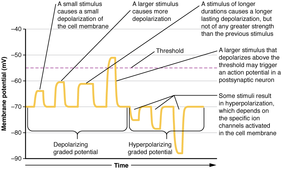
Graded potentials are temporary changes in the membrane voltage, the characteristics of which depend on the size of the stimulus. Some types of stimuli cause depolarization of the membrane, whereas others cause hyperpolarization. It depends on the specific ion channels that are activated in the cell membrane.

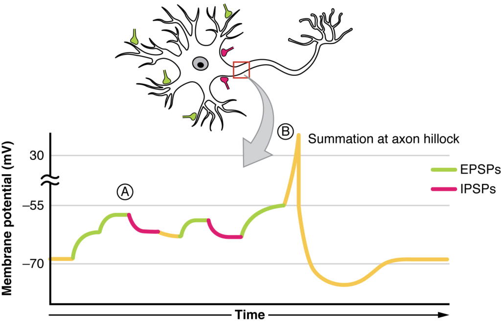
The result of summation of postsynaptic potentials is the overall change in the membrane potential. At point A, several different excitatory postsynaptic potentials add up to a large depolarization. At point B, a mix of excitatory and inhibitory postsynaptic potentials result in a different end result for the membrane potential.

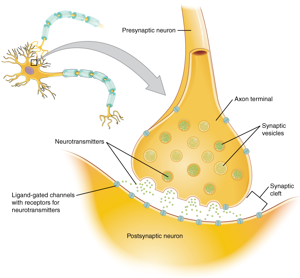
The synapse is a connection between a neuron and its target cell (which is not necessarily a neuron). The presynaptic element is the synaptic end bulb of the axon where Ca2+ enters the bulb to cause vesicle fusion and neurotransmitter release. The neurotransmitter diffuses across the synaptic cleft to bind to its receptor. The neurotransmitter is cleared from the synapse either by enzymatic degradation, neuronal reuptake, or glial reuptake.

(a) An ionotropic receptor is a channel that opens when the neurotransmitter binds to it. (b) A metabotropic receptor is a complex that causes metabolic changes in the cell when the neurotransmitter binds to it (1). After binding, the G protein hydrolyzes ATP and moves to the effector protein (2). When the G protein contacts the effector protein, the latter is activated. In the case shown, the effector protein then acts on ATP to generate a second messenger, cAMP (3). The second messenger can then go on to cause changes in the neuron, such as opening or closing ion channels, metabolic changes, and changes in gene transcription.

SystemCholinergicAmino acidsBiogenic aminesNeuropeptides
Neuro-transmittersAcetylcholineGlutamate, glycine, GABASerotonin (5-HT), dopamine, norepinephrine, (epinephrine)Met-enkephalin, beta-endorphin, VIP, Substance P, etc.
ReceptorsNicotinic and muscarinic receptorsGlu receptors, gly receptors, GABA receptors5-HT receptors, D1 and D2 receptors, α-adrenergic and β-adrenergic receptorsReceptors are too numerous to list, but are specific to the peptides.
EliminationDegradation by acetylcholinesteraseReuptake by neurons or gliaReuptake by neuronsDegradation by enzymes called peptidases
Postsynaptic effectNicotinic receptor causes depolarization. Muscarinic receptors can cause both depolarization or hyperpolarization depending on the subtype.Glu receptors cause depolarization. Gly and GABA receptors cause hyperpolarization.Depolarization or hyperpolarization depends on the specific receptor. For example, D1 receptors cause depolarization and D2 receptors cause hyperpolarization.Depolarization or hyperpolarization depends on the specific receptor.

Nội dung này đang được cập nhật.
Dưới đây là video và các luyện tập (practice) của bài này. Nghe là một kĩ năng khó, đặc biệt là khi chúng ta chưa quen nội dung và chưa có nhạy cảm ngôn ngữ. Nhưng cứ đi thật chậm và đừng bỏ cuộc.
Xem video và cảm nhận nội dung bài. Bạn có thể thả trôi, cảm nhận dòng chảy ngôn ngữ và không nhất thiết phải hiểu toàn bộ bài. Bên dưới là script để bạn khái quát nội dụng và tra từ mới.
Script:
  1. The basis of the electrical signal within a neuron is the action potential that propagates down the axon.
  2. For a neuron to generate an action potential, it needs to receive input from another source, either another neuron or a sensory stimulus.
  3. That input will result in opening ion channels in the neuron, resulting in a graded potential based on the strength of the stimulus.
  4. Graded potentials can be depolarizing or hyperpolarizing and can summate to affect the probability of the neuron reaching threshold.
  5. Graded potentials can be the result of sensory stimuli.
  6. If the sensory stimulus is received by the dendrites of a unipolar sensory neuron, such as the sensory neuron ending in the skin, the graded potential is called a generator potential because it can directly generate the action potential in the initial segment of the axon.
  7. If the sensory stimulus is received by a specialized sensory receptor cell, the graded potential is called a receptor potential.
  8. Graded potentials produced by interactions between neurons at synapses are called postsynaptic potentials.
  9. A depolarizing graded potential at a synapse is called an excitatory postsynaptic potential, and a hyperpolarizing graded potential at a synapse is called an inhibitory postsynaptic potential.
  10. Synapses are the contacts between neurons, which can either be chemical or electrical in nature.
  11. Chemical synapses are far more common.
  12. At a chemical synapse, neurotransmitter is released from the presynaptic element and diffuses across the synaptic cleft.
  13. The neurotransmitter binds to a receptor protein and causes a change in the postsynaptic membrane (the postsynaptic potential).
  14. The neurotransmitter must be inactivated or removed from the synaptic cleft so that the stimulus is limited in time.
  15. The particular characteristics of a synapse vary based on the neurotransmitter system produced by that neuron.
  16. The cholinergic system is found at the neuromuscular junction and in certain places within the nervous system.
  17. Amino acids, such as glutamate, glycine, and gamma-aminobutyric acid are used as neurotransmitters.
  18. Other neurotransmitters are the result of amino acids being enzymatically changed, as in the biogenic amines, or being covalently bonded together, as in the neuropeptides.
Bật video, nghe và điền từ vào chỗ trống.
Dưới đây là phần bàn luận. Bạn có thể tự do đặt câu hỏi, bổ sung kiến thức, và chia sẻ trải nghiệm của mình.
Subscribe
Notify of

0 Comments
Inline Feedbacks
View all comments

Ấn vào ô bên dưới để đánh dấu bạn đã hoàn thành bài học này

Quá dữ! Tiếp tục duy trì phong độ nhé!