Module 8: Anatomy of the Nervous System

Lesson 4: The Peripheral Nervous System

Hệ Thần Kinh Ngoại Biên

Nội dung bài học:
Mỗi bài học (lesson) bao gồm 4 phần chính: Thuật ngữ, Luyện Đọc, Luyện Nghe, và Bàn Luận.
Sử dụng tính năng:
Bôi hoặc nhấp đôi vào từ, sau đó ấn vào biểu tượng để tra nghĩa từ đó. Khi bạn đưa chuột đến câu (hoặc khi nhấp vào câu trên màn hình điện thoại), gợi ý về cách hiểu câu đó sẽ hiện lên. Cuối cùng, bạn có thể đánh dấu hoàn thành toàn bộ bài học bằng cách ấn vào nút “Hoàn Thành” ở cuối trang.
Đăng ký và đăng nhập
Bạn cần đăng ký và đăng nhập vào tài khoản để lưu quá trình học.
Dưới đây là danh sách những thuật ngữ Y khoa của module Anatomy of the Nervous System.
Khái quát được số lượng thuật ngữ sẽ xuất hiện trong bài đọc và nghe sẽ giúp bạn thoải mái tiêu thụ nội dung hơn. Sau khi hoàn thành nội dung đọc và nghe, bạn hãy quay lại đây và luyện tập (practice) để quen dần các thuật ngữ này. Đừng ép bản thân phải nhớ các thuật ngữ này vội vì bạn sẽ gặp và ôn lại danh sách này trong những bài học (lesson) khác của cùng một module.

Medical Terminology: Anatomy of the Nervous System

abducens nerve
sixth cranial nerve; responsible for contraction of one of the extraocular muscles
alar plate
developmental region of the spinal cord that gives rise to the posterior horn of the gray matter
amygdala
nucleus deep in the temporal lobe of the cerebrum that is related to memory and emotional behavior
anterior column
white matter between the anterior horns of the spinal cord composed of many different groups of axons of both ascending and descending tracts
anterior horn
gray matter of the spinal cord containing multipolar motor neurons, sometimes referred to as the ventral horn
anterior median fissure
deep midline feature of the anterior spinal cord, marking the separation between the right and left sides of the cord
anterior spinal artery
blood vessel from the merged branches of the vertebral arteries that runs along the anterior surface of the spinal cord
arachnoid granulation
outpocket of the arachnoid membrane into the dural sinuses that allows for reabsorption of CSF into the blood
arachnoid mater
middle layer of the meninges named for the spider-web–like trabeculae that extend between it and the pia mater
arachnoid trabeculae
filaments between the arachnoid and pia mater within the subarachnoid space
ascending tract
central nervous system fibers carrying sensory information from the spinal cord or periphery to the brain
axillary nerve
systemic nerve of the arm that arises from the brachial plexus
basal forebrain
nuclei of the cerebrum related to modulation of sensory stimuli and attention through broad projections to the cerebral cortex, loss of which is related to Alzheimer’s disease
basal nuclei
nuclei of the cerebrum (with a few components in the upper brain stem and diencephalon) that are responsible for assessing cortical movement commands and comparing them with the general state of the individual through broad modulatory activity of dopamine neurons; largely related to motor functions, as evidenced through the symptoms of Parkinson’s and Huntington’s diseases
basal plate
developmental region of the spinal cord that gives rise to the lateral and anterior horns of gray matter
basilar artery
blood vessel from the merged vertebral arteries that runs along the dorsal surface of the brain stem
brachial plexus
nerve plexus associated with the lower cervical spinal nerves and first thoracic spinal nerve
brain stem
region of the adult brain that includes the midbrain, pons, and medulla oblongata and develops from the mesencephalon, metencephalon, and myelencephalon of the embryonic brain
Broca’s area
region of the frontal lobe associated with the motor commands necessary for speech production and located only in the cerebral hemisphere responsible for language production, which is the left side in approximately 95 percent of the population
Brodmann’s areas
mapping of regions of the cerebral cortex based on microscopic anatomy that relates specific areas to functional differences, as described by Brodmann in the early 1900s
carotid canal
opening in the temporal bone through which the internal carotid artery enters the cranium
cauda equina
bundle of spinal nerve roots that descend from the lower spinal cord below the first lumbar vertebra and lie within the vertebral cavity; has the appearance of a horse’s tail
caudate
nucleus deep in the cerebrum that is part of the basal nuclei; along with the putamen, it is part of the striatum
central canal
hollow space within the spinal cord that is the remnant of the center of the neural tube
central sulcus
surface landmark of the cerebral cortex that marks the boundary between the frontal and parietal lobes
cephalic flexure
curve in midbrain of the embryo that positions the forebrain ventrally
cerebellum
region of the adult brain connected primarily to the pons that developed from the metencephalon (along with the pons) and is largely responsible for comparing information from the cerebrum with sensory feedback from the periphery through the spinal cord
cerebral aqueduct
connection of the ventricular system between the third and fourth ventricles located in the midbrain
cerebral cortex
outer gray matter covering the forebrain, marked by wrinkles and folds known as gyri and sulci
cerebral hemisphere
one half of the bilaterally symmetrical cerebrum
cerebrum
region of the adult brain that develops from the telencephalon and is responsible for higher neurological functions such as memory, emotion, and consciousness
cervical plexus
nerve plexus associated with the upper cervical spinal nerves
choroid plexus
specialized structures containing ependymal cells lining blood capillaries that filter blood to produce CSF in the four ventricles of the brain
circle of Willis
unique anatomical arrangement of blood vessels around the base of the brain that maintains perfusion of blood into the brain even if one component of the structure is blocked or narrowed
common carotid artery
blood vessel that branches off the aorta (or the brachiocephalic artery on the right) and supplies blood to the head and neck
corpus callosum
large white matter structure that connects the right and left cerebral hemispheres
cranial nerve
one of twelve nerves connected to the brain that are responsible for sensory or motor functions of the head and neck
cranial nerve ganglion
sensory ganglion of cranial nerves
descending tract
central nervous system fibers carrying motor commands from the brain to the spinal cord or periphery
diencephalon
region of the adult brain that retains its name from embryonic development and includes the thalamus and hypothalamus
direct pathway
connections within the basal nuclei from the striatum to the globus pallidus internal segment and substantia nigra pars reticulata that disinhibit the thalamus to increase cortical control of movement
disinhibition
disynaptic connection in which the first synapse inhibits the second cell, which then stops inhibiting the final target
dorsal (posterior) nerve root
axons entering the posterior horn of the spinal cord
dorsal (posterior) root ganglion
sensory ganglion attached to the posterior nerve root of a spinal nerve
dura mater
tough, fibrous, outer layer of the meninges that is attached to the inner surface of the cranium and vertebral column and surrounds the entire CNS
dural sinus
any of the venous structures surrounding the brain, enclosed within the dura mater, which drain blood from the CNS to the common venous return of the jugular veins
endoneurium
innermost layer of connective tissue that surrounds individual axons within a nerve
enteric nervous system
peripheral structures, namely ganglia and nerves, that are incorporated into the digestive system organs
enteric plexus
neuronal plexus in the wall of the intestines, which is part of the enteric nervous system
epineurium
outermost layer of connective tissue that surrounds an entire nerve
epithalamus
region of the diecephalon containing the pineal gland
esophageal plexus
neuronal plexus in the wall of the esophagus that is part of the enteric nervous system
extraocular muscles
six skeletal muscles that control eye movement within the orbit
facial nerve
seventh cranial nerve; responsible for contraction of the facial muscles and for part of the sense of taste, as well as causing saliva production
fascicle
small bundles of nerve or muscle fibers enclosed by connective tissue
femoral nerve
systemic nerve of the anterior leg that arises from the lumbar plexus
fibular nerve
systemic nerve of the posterior leg that begins as part of the sciatic nerve
foramen magnum
large opening in the occipital bone of the skull through which the spinal cord emerges and the vertebral arteries enter the cranium
forebrain
anterior region of the adult brain that develops from the prosencephalon and includes the cerebrum and diencephalon
fourth ventricle
the portion of the ventricular system that is in the region of the brain stem and opens into the subarachnoid space through the median and lateral apertures
frontal eye field
region of the frontal lobe associated with motor commands to orient the eyes toward an object of visual attention
frontal lobe
region of the cerebral cortex directly beneath the frontal bone of the cranium
gastric plexuses
neuronal networks in the wall of the stomach that are part of the enteric nervous system
globus pallidus
nuclei deep in the cerebrum that are part of the basal nuclei and can be divided into the internal and external segments
glossopharyngeal nerve
ninth cranial nerve; responsible for contraction of muscles in the tongue and throat and for part of the sense of taste, as well as causing saliva production
gyrus
ridge formed by convolutions on the surface of the cerebrum or cerebellum
hindbrain
posterior region of the adult brain that develops from the rhombencephalon and includes the pons, medulla oblongata, and cerebellum
hippocampus
gray matter deep in the temporal lobe that is very important for long-term memory formation
hypoglossal nerve
twelfth cranial nerve; responsible for contraction of muscles of the tongue
hypothalamus
major region of the diencephalon that is responsible for coordinating autonomic and endocrine control of homeostasis
indirect pathway
connections within the basal nuclei from the striatum through the globus pallidus external segment and subthalamic nucleus to the globus pallidus internal segment/substantia nigra pars compacta that result in inhibition of the thalamus to decrease cortical control of movement
inferior colliculus
half of the midbrain tectum that is part of the brain stem auditory pathway
inferior olive
nucleus in the medulla that is involved in processing information related to motor control
intercostal nerve
systemic nerve in the thoracic cavity that is found between two ribs
internal carotid artery
branch from the common carotid artery that enters the cranium and supplies blood to the brain
interventricular foramina
openings between the lateral ventricles and third ventricle allowing for the passage of CSF
jugular veins
blood vessels that return “used” blood from the head and neck
kinesthesia
general sensory perception of movement of the body
lateral apertures
pair of openings from the fourth ventricle to the subarachnoid space on either side and between the medulla and cerebellum
lateral column
white matter of the spinal cord between the posterior horn on one side and the axons from the anterior horn on the same side; composed of many different groups of axons, of both ascending and descending tracts, carrying motor commands to and from the brain
lateral horn
region of the spinal cord gray matter in the thoracic, upper lumbar, and sacral regions that is the central component of the sympathetic division of the autonomic nervous system
lateral sulcus
surface landmark of the cerebral cortex that marks the boundary between the temporal lobe and the frontal and parietal lobes
lateral ventricles
portions of the ventricular system that are in the region of the cerebrum
limbic cortex
collection of structures of the cerebral cortex that are involved in emotion, memory, and behavior and are part of the larger limbic system
limbic system
structures at the edge (limit) of the boundary between the forebrain and hindbrain that are most associated with emotional behavior and memory formation
longitudinal fissure
large separation along the midline between the two cerebral hemispheres
lumbar plexus
nerve plexus associated with the lumbar spinal nerves
lumbar puncture
procedure used to withdraw CSF from the lower lumbar region of the vertebral column that avoids the risk of damaging CNS tissue because the spinal cord ends at the upper lumbar vertebrae
median aperture
singular opening from the fourth ventricle into the subarachnoid space at the midline between the medulla and cerebellum
median nerve
systemic nerve of the arm, located between the ulnar and radial nerves
meninges
protective outer coverings of the CNS composed of connective tissue
mesencephalon
primary vesicle of the embryonic brain that does not significantly change through the rest of embryonic development and becomes the midbrain
metencephalon
secondary vesicle of the embryonic brain that develops into the pons and the cerebellum
midbrain
middle region of the adult brain that develops from the mesencephalon
myelencephalon
secondary vesicle of the embryonic brain that develops into the medulla
nerve plexus
network of nerves without neuronal cell bodies included
neural crest
tissue that detaches from the edges of the neural groove and migrates through the embryo to develop into peripheral structures of both nervous and non-nervous tissues
neural fold
elevated edge of the neural groove
neural groove
region of the neural plate that folds into the dorsal surface of the embryo and closes off to become the neural tube
neural plate
thickened layer of neuroepithelium that runs longitudinally along the dorsal surface of an embryo and gives rise to nervous system tissue
neural tube
precursor to structures of the central nervous system, formed by the invagination and separation of neuroepithelium
neuraxis
central axis to the nervous system, from the posterior to anterior ends of the neural tube; the inferior tip of the spinal cord to the anterior surface of the cerebrum
occipital lobe
region of the cerebral cortex directly beneath the occipital bone of the cranium
occipital sinuses
dural sinuses along the edge of the occipital lobes of the cerebrum
oculomotor nerve
third cranial nerve; responsible for contraction of four of the extraocular muscles, the muscle in the upper eyelid, and pupillary constriction
olfaction
special sense responsible for smell, which has a unique, direct connection to the cerebrum
olfactory nerve
first cranial nerve; responsible for the sense of smell
optic nerve
second cranial nerve; responsible for visual sensation
orthostatic reflex
sympathetic function that maintains blood pressure when standing to offset the increased effect of gravity
paravertebral ganglia
autonomic ganglia superior to the sympathetic chain ganglia
parietal lobe
region of the cerebral cortex directly beneath the parietal bone of the cranium
parieto-occipital sulcus
groove in the cerebral cortex representing the border between the parietal and occipital cortices
perineurium
layer of connective tissue surrounding fascicles within a nerve
phrenic nerve
systemic nerve from the cervical plexus that innervates the diaphragm
pia mater
thin, innermost membrane of the meninges that directly covers the surface of the CNS
plexus
network of nerves or nervous tissue
postcentral gyrus
primary motor cortex located in the frontal lobe of the cerebral cortex
posterior columns
white matter of the spinal cord that lies between the posterior horns of the gray matter, sometimes referred to as the dorsal column; composed of axons of ascending tracts that carry sensory information up to the brain
posterior horn
gray matter region of the spinal cord in which sensory input arrives, sometimes referred to as the dorsal horn
posterior median sulcus
midline feature of the posterior spinal cord, marking the separation between right and left sides of the cord
posterolateral sulcus
feature of the posterior spinal cord marking the entry of posterior nerve roots and the separation between the posterior and lateral columns of the white matter
precentral gyrus
ridge just posterior to the central sulcus, in the parietal lobe, where somatosensory processing initially takes place in the cerebrum
prefrontal lobe
specific region of the frontal lobe anterior to the more specific motor function areas, which can be related to the early planning of movements and intentions to the point of being personality-type functions
premotor area
region of the frontal lobe responsible for planning movements that will be executed through the primary motor cortex
prevertebral ganglia
autonomic ganglia that are anterior to the vertebral column and functionally related to the sympathetic chain ganglia
primary vesicle
initial enlargements of the anterior neural tube during embryonic development that develop into the forebrain, midbrain, and hindbrain
proprioception
general sensory perceptions providing information about location and movement of body parts; the “sense of the self”
prosencephalon
primary vesicle of the embryonic brain that develops into the forebrain, which includes the cerebrum and diencephalon
putamen
nucleus deep in the cerebrum that is part of the basal nuclei; along with the caudate, it is part of the striatum
radial nerve
systemic nerve of the arm, the distal component of which is located near the radial bone
reticular formation
diffuse region of gray matter throughout the brain stem that regulates sleep, wakefulness, and states of consciousness
rhombencephalon
primary vesicle of the embryonic brain that develops into the hindbrain, which includes the pons, cerebellum, and medulla
sacral plexus
nerve plexus associated with the lower lumbar and sacral spinal nerves
saphenous nerve
systemic nerve of the lower anterior leg that is a branch from the femoral nerve
sciatic nerve
systemic nerve from the sacral plexus that is a combination of the tibial and fibular nerves and extends across the hip joint and gluteal region into the upper posterior leg
sciatica
painful condition resulting from inflammation or compression of the sciatic nerve or any of the spinal nerves that contribute to it
secondary vesicle
five vesicles that develop from primary vesicles, continuing the process of differentiation of the embryonic brain
sigmoid sinuses
dural sinuses that drain directly into the jugular veins
somatosensation
general senses related to the body, usually thought of as the senses of touch, which would include pain, temperature, and proprioception
spinal accessory nerve
eleventh cranial nerve; responsible for contraction of neck muscles
spinal nerve
one of 31 nerves connected to the spinal cord
straight sinus
dural sinus that drains blood from the deep center of the brain to collect with the other sinuses
striatum
the caudate and putamen collectively, as part of the basal nuclei, which receive input from the cerebral cortex
subarachnoid space
space between the arachnoid mater and pia mater that contains CSF and the fibrous connections of the arachnoid trabeculae
subcortical nucleus
all the nuclei beneath the cerebral cortex, including the basal nuclei and the basal forebrain
substantia nigra pars compacta
nuclei within the basal nuclei that release dopamine to modulate the function of the striatum; part of the motor pathway
substantia nigra pars reticulata
nuclei within the basal nuclei that serve as an output center of the nuclei; part of the motor pathway
subthalamus
nucleus within the basal nuclei that is part of the indirect pathway
sulcus
groove formed by convolutions in the surface of the cerebral cortex
superior colliculus
half of the midbrain tectum that is responsible for aligning visual, auditory, and somatosensory spatial perceptions
superior sagittal sinus
dural sinus that runs along the top of the longitudinal fissure and drains blood from the majority of the outer cerebrum
sympathetic chain ganglia
autonomic ganglia in a chain along the anterolateral aspect of the vertebral column that are responsible for contributing to homeostatic mechanisms of the autonomic nervous system
systemic nerve
nerve in the periphery distal to a nerve plexus or spinal nerve
tectum
region of the midbrain, thought of as the roof of the cerebral aqueduct, which is subdivided into the inferior and superior colliculi
tegmentum
region of the midbrain, thought of as the floor of the cerebral aqueduct, which continues into the pons and medulla as the floor of the fourth ventricle
telencephalon
secondary vesicle of the embryonic brain that develops into the cerebrum
temporal lobe
region of the cerebral cortex directly beneath the temporal bone of the cranium
terminal ganglion
autonomic ganglia that are near or within the walls of organs that are responsible for contributing to homeostatic mechanisms of the autonomic nervous system
thalamus
major region of the diencephalon that is responsible for relaying information between the cerebrum and the hindbrain, spinal cord, and periphery
third ventricle
portion of the ventricular system that is in the region of the diencephalon
tibial nerve
systemic nerve of the posterior leg that begins as part of the sciatic nerve
transverse sinuses
dural sinuses that drain along either side of the occipital–cerebellar space
trigeminal ganglion
sensory ganglion that contributes sensory fibers to the trigeminal nerve
trigeminal nerve
fifth cranial nerve; responsible for cutaneous sensation of the face and contraction of the muscles of mastication
trochlear nerve
fourth cranial nerve; responsible for contraction of one of the extraocular muscles
ulnar nerve
systemic nerve of the arm located close to the ulna, a bone of the forearm
vagus nerve
tenth cranial nerve; responsible for the autonomic control of organs in the thoracic and upper abdominal cavities
ventral (anterior) nerve root
axons emerging from the anterior or lateral horns of the spinal cord
ventricles
remnants of the hollow center of the neural tube that are spaces for cerebrospinal fluid to circulate through the brain
vertebral arteries
arteries that ascend along either side of the vertebral column through the transverse foramina of the cervical vertebrae and enter the cranium through the foramen magnum
vestibulocochlear nerve
eighth cranial nerve; responsible for the sensations of hearing and balance
Nội dung này đang được cập nhật.
Dưới đây là các bài văn nằm ở bên trái. Ở bên phải là các bài luyện tập (practice) để đánh giá khả năng đọc hiểu của bạn. Sẽ khó khăn trong thời gian đầu nếu vốn từ vựng của bạn còn hạn chế, đặc biệt là từ vựng Y khoa. Hãy kiên nhẫn và đọc nhiều nhất có kể, lượng kiến thức tích tụ dần sẽ giúp bạn đọc thoải mái hơn.
The PNS is not as contained as the CNS because it is defined as everything that is not the CNS. Some peripheral structures are incorporated into the other organs of the body. In describing the anatomy of the PNS, it is necessary to describe the common structures, the nerves and the ganglia, as they are found in various parts of the body. Many of the neural structures that are incorporated into other organs are features of the digestive system; these structures are known as the enteric nervous system and are a special subset of the PNS.

A ganglion is a group of neuron cell bodies in the periphery. Ganglia can be categorized, for the most part, as either sensory ganglia or autonomic ganglia, referring to their primary functions. The most common type of sensory ganglion is a dorsal (posterior) root ganglion. These ganglia are the cell bodies of neurons with axons that are sensory endings in the periphery, such as in the skin, and that extend into the CNS through the dorsal nerve root. The ganglion is an enlargement of the nerve root. Under microscopic inspection, it can be seen to include the cell bodies of the neurons, as well as bundles of fibers that are the posterior nerve root (Figure 1 and Figure 2). The cells of the dorsal root ganglion are unipolar cells, classifying them by shape. Also, the small round nuclei of satellite cells can be seen surrounding—as if they were orbiting—the neuron cell bodies.

Another type of sensory ganglion is a cranial nerve ganglion. This is analogous to the dorsal root ganglion, except that it is associated with a cranial nerve instead of a spinal nerve. The roots of cranial nerves are within the cranium, whereas the ganglia are outside the skull. For example, the trigeminal ganglion is superficial to the temporal bone whereas its associated nerve is attached to the mid-pons region of the brain stem. The neurons of cranial nerve ganglia are also unipolar in shape with associated satellite cells.

The other major category of ganglia are those of the autonomic nervous system, which is divided into the sympathetic and parasympathetic nervous systems. The sympathetic chain ganglia constitute a row of ganglia along the vertebral column that receive central input from the lateral horn of the thoracic and upper lumbar spinal cord. Superior to the chain ganglia are three paravertebral ganglia in the cervical region. Three other autonomic ganglia that are related to the sympathetic chain are the prevertebral ganglia, which are located outside of the chain but have similar functions. They are referred to as prevertebral because they are anterior to the vertebral column. The neurons of these autonomic ganglia are multipolar in shape, with dendrites radiating out around the cell body where synapses from the spinal cord neurons are made. The neurons of the chain, paravertebral, and prevertebral ganglia then project to organs in the head and neck, thoracic, abdominal, and pelvic cavities to regulate the sympathetic aspect of homeostatic mechanisms.

Another group of autonomic ganglia are the terminal ganglia that receive input from cranial nerves or sacral spinal nerves and are responsible for regulating the parasympathetic aspect of homeostatic mechanisms. These two sets of ganglia, sympathetic and parasympathetic, often project to the same organs—one input from the chain ganglia and one input from a terminal ganglion—to regulate the overall function of an organ. For example, the heart receives two inputs such as these; one increases heart rate, and the other decreases it. The terminal ganglia that receive input from cranial nerves are found in the head and neck, as well as the thoracic and upper abdominal cavities, whereas the terminal ganglia that receive sacral input are in the lower abdominal and pelvic cavities.

Terminal ganglia below the head and neck are often incorporated into the wall of the target organ as a plexus. A plexus, in a general sense, is a network of fibers or vessels. This can apply to nervous tissue (as in this instance) or structures containing blood vessels (such as a choroid plexus). For example, the enteric plexus is the extensive network of axons and neurons in the wall of the small and large intestines. The enteric plexus is actually part of the enteric nervous system, along with the gastric plexuses and the esophageal plexus. Though the enteric nervous system receives input originating from central neurons of the autonomic nervous system, it does not require CNS input to function. In fact, it operates independently to regulate the digestive system.
Bundles of axons in the PNS are referred to as nerves. These structures in the periphery are different than the central counterpart, called a tract. Nerves are composed of more than just nervous tissue. They have connective tissues invested in their structure, as well as blood vessels supplying the tissues with nourishment. The outer surface of a nerve is a surrounding layer of fibrous connective tissue called the epineurium. Within the nerve, axons are further bundled into fascicles, which are each surrounded by their own layer of fibrous connective tissue called perineurium. Finally, individual axons are surrounded by loose connective tissue called the endoneurium (Figure 3 and Figure 4). These three layers are similar to the connective tissue sheaths for muscles. Nerves are associated with the region of the CNS to which they are connected, either as cranial nerves connected to the brain or spinal nerves connected to the spinal cord.

A. Cranial Nerves

The nerves attached to the brain are the cranial nerves, which are primarily responsible for the sensory and motor functions of the head and neck (one of these nerves targets organs in the thoracic and abdominal cavities as part of the parasympathetic nervous system). There are twelve cranial nerves, which are designated CNI through CNXII for “Cranial Nerve,” using Roman numerals for 1 through 12. They can be classified as sensory nerves, motor nerves, or a combination of both, meaning that the axons in these nerves originate out of sensory ganglia external to the cranium or motor nuclei within the brain stem. Sensory axons enter the brain to synapse in a nucleus. Motor axons connect to skeletal muscles of the head or neck. Three of the nerves are solely composed of sensory fibers; five are strictly motor; and the remaining four are mixed nerves.

Learning the cranial nerves is a tradition in anatomy courses, and students have always used mnemonic devices to remember the nerve names. A traditional mnemonic is the rhyming couplet, “On Old Olympus’ Towering Tops/A Finn And German Viewed Some Hops,” in which the initial letter of each word corresponds to the initial letter in the name of each nerve. The names of the nerves have changed over the years to reflect current usage and more accurate naming. An exercise to help learn this sort of information is to generate a mnemonic using words that have personal significance. The names of the cranial nerves are listed in Table 1 along with a brief description of their function, their source (sensory ganglion or motor nucleus), and their target (sensory nucleus or skeletal muscle). They are listed here with a brief explanation of each nerve (Figure 5).

The olfactory nerve and optic nerve are responsible for the sense of smell and vision, respectively. The oculomotor nerve is responsible for eye movements by controlling four of the extraocular muscles. It is also responsible for lifting the upper eyelid when the eyes point up, and for pupillary constriction. The trochlear nerve and the abducens nerve are both responsible for eye movement, but do so by controlling different extraocular muscles. The trigeminal nerve is responsible for cutaneous sensations of the face and controlling the muscles of mastication. The facial nerve is responsible for the muscles involved in facial expressions, as well as part of the sense of taste and the production of saliva. The vestibulocochlear nerve is responsible for the senses of hearing and balance. The glossopharyngeal nerve is responsible for controlling muscles in the oral cavity and upper throat, as well as part of the sense of taste and the production of saliva. The vagus nerve is responsible for contributing to homeostatic control of the organs of the thoracic and upper abdominal cavities. The spinal accessory nerve is responsible for controlling the muscles of the neck, along with cervical spinal nerves. The hypoglossal nerve is responsible for controlling the muscles of the lower throat and tongue.

Three of the cranial nerves also contain autonomic fibers, and a fourth is almost purely a component of the autonomic system. The oculomotor, facial, and glossopharyngeal nerves contain fibers that contact autonomic ganglia. The oculomotor fibers initiate pupillary constriction, whereas the facial and glossopharyngeal fibers both initiate salivation. The vagus nerve primarily targets autonomic ganglia in the thoracic and upper abdominal cavities.

Another important aspect of the cranial nerves that lends itself to a mnemonic is the functional role each nerve plays. The nerves fall into one of three basic groups. They are sensory, motor, or both (see Table 13.3). The sentence, “Some Say Marry Money But My Brother Says Brains Beauty Matter More,” corresponds to the basic function of each nerve. The first, second, and eighth nerves are purely sensory: the olfactory (CNI), optic (CNII), and vestibulocochlear (CNVIII) nerves. The three eye-movement nerves are all motor: the oculomotor (CNIII), trochlear (CNIV), and abducens (CNVI). The spinal accessory (CNXI) and hypoglossal (CNXII) nerves are also strictly motor. The remainder of the nerves contain both sensory and motor fibers. They are the trigeminal (CNV), facial (CNVII), glossopharyngeal (CNIX), and vagus (CNX) nerves. The nerves that convey both are often related to each other. The trigeminal and facial nerves both concern the face; one concerns the sensations and the other concerns the muscle movements. The facial and glossopharyngeal nerves are both responsible for conveying gustatory, or taste, sensations as well as controlling salivary glands. The vagus nerve is involved in visceral responses to taste, namely the gag reflex. This is not an exhaustive list of what these combination nerves do, but there is a thread of relation between them.

B. Spinal Nerves

The nerves connected to the spinal cord are the spinal nerves. The arrangement of these nerves is much more regular than that of the cranial nerves. All of the spinal nerves are combined sensory and motor axons that separate into two nerve roots. The sensory axons enter the spinal cord as the dorsal nerve root. The motor fibers, both somatic and autonomic, emerge as the ventral nerve root. The dorsal root ganglion for each nerve is an enlargement of the spinal nerve.

There are 31 spinal nerves, named for the level of the spinal cord at which each one emerges. There are eight pairs of cervical nerves designated C1 to C8, twelve thoracic nerves designated T1 to T12, five pairs of lumbar nerves designated L1 to L5, five pairs of sacral nerves designated S1 to S5, and one pair of coccygeal nerves. The nerves are numbered from the superior to inferior positions, and each emerges from the vertebral column through the intervertebral foramen at its level. The first nerve, C1, emerges between the first cervical vertebra and the occipital bone. The second nerve, C2, emerges between the first and second cervical vertebrae. The same occurs for C3 to C7, but C8 emerges between the seventh cervical vertebra and the first thoracic vertebra. For the thoracic and lumbar nerves, each one emerges between the vertebra that has the same designation and the next vertebra in the column. The sacral nerves emerge from the sacral foramina along the length of that unique vertebra.

Spinal nerves extend outward from the vertebral column to innervate the periphery. The nerves in the periphery are not straight continuations of the spinal nerves, but rather the reorganization of the axons in those nerves to follow different courses. Axons from different spinal nerves will come together into a systemic nerve. This occurs at four places along the length of the vertebral column, each identified as a nerve plexus, whereas the other spinal nerves directly correspond to nerves at their respective levels. In this instance, the word plexus is used to describe networks of nerve fibers with no associated cell bodies.

Of the four nerve plexuses, two are found at the cervical level, one at the lumbar level, and one at the sacral level (Figure 6). The cervical plexus is composed of axons from spinal nerves C1 through C5 and branches into nerves in the posterior neck and head, as well as the phrenic nerve, which connects to the diaphragm at the base of the thoracic cavity. The other plexus from the cervical level is the brachial plexus. Spinal nerves C4 through T1 reorganize through this plexus to give rise to the nerves of the arms, as the name brachial suggests. A large nerve from this plexus is the radial nerve from which the axillary nerve branches to go to the armpit region. The radial nerve continues through the arm and is paralleled by the ulnar nerve and the median nerve. The lumbar plexus arises from all the lumbar spinal nerves and gives rise to nerves enervating the pelvic region and the anterior leg. The femoral nerve is one of the major nerves from this plexus, which gives rise to the saphenous nerve as a branch that extends through the anterior lower leg. The sacral plexus comes from the lower lumbar nerves L4 and L5 and the sacral nerves S1 to S4. The most significant systemic nerve to come from this plexus is the sciatic nerve, which is a combination of the tibial nerve and the fibular nerve. The sciatic nerve extends across the hip joint and is most commonly associated with the condition sciatica, which is the result of compression or irritation of the nerve or any of the spinal nerves giving rise to it.

These plexuses are described as arising from spinal nerves and giving rise to certain systemic nerves, but they contain fibers that serve sensory functions or fibers that serve motor functions. This means that some fibers extend from cutaneous or other peripheral sensory surfaces and send action potentials into the CNS. Those are axons of sensory neurons in the dorsal root ganglia that enter the spinal cord through the dorsal nerve root. Other fibers are the axons of motor neurons of the anterior horn of the spinal cord, which emerge in the ventral nerve root and send action potentials to cause skeletal muscles to contract in their target regions. For example, the radial nerve contains fibers of cutaneous sensation in the arm, as well as motor fibers that move muscles in the arm.

Spinal nerves of the thoracic region, T2 through T11, are not part of the plexuses but rather emerge and give rise to the intercostal nerves found between the ribs, which articulate with the vertebrae surrounding the spinal nerve.

OpenStax. (2022). Anatomy and Physiology 2e. Rice University. Retrieved June 15, 2023. ISBN-13: 978-1-711494-06-7 (Hardcover) ISBN-13: 978-1-711494-05-0 (Paperback) ISBN-13: 978-1-951693-42-8 (Digital). License: Attribution 4.0 International (CC BY 4.0). Access for free at openstax.org.

The cell bodies of sensory neurons, which are unipolar neurons by shape, are seen in this photomicrograph. Also, the fibrous region is composed of the axons of these neurons that are passing through the ganglion to be part of the dorsal nerve root (tissue source: canine). LM × 40. (Micrograph provided by the Regents of University of Michigan Medical School © 2012)

The slide includes both a cross-section of the lumbar spinal cord and a section of the dorsal root ganglion (see also Figure 13.19) (tissue source: canine). LM × 1600. (Micrograph provided by the Regents of University of Michigan Medical School © 2012)

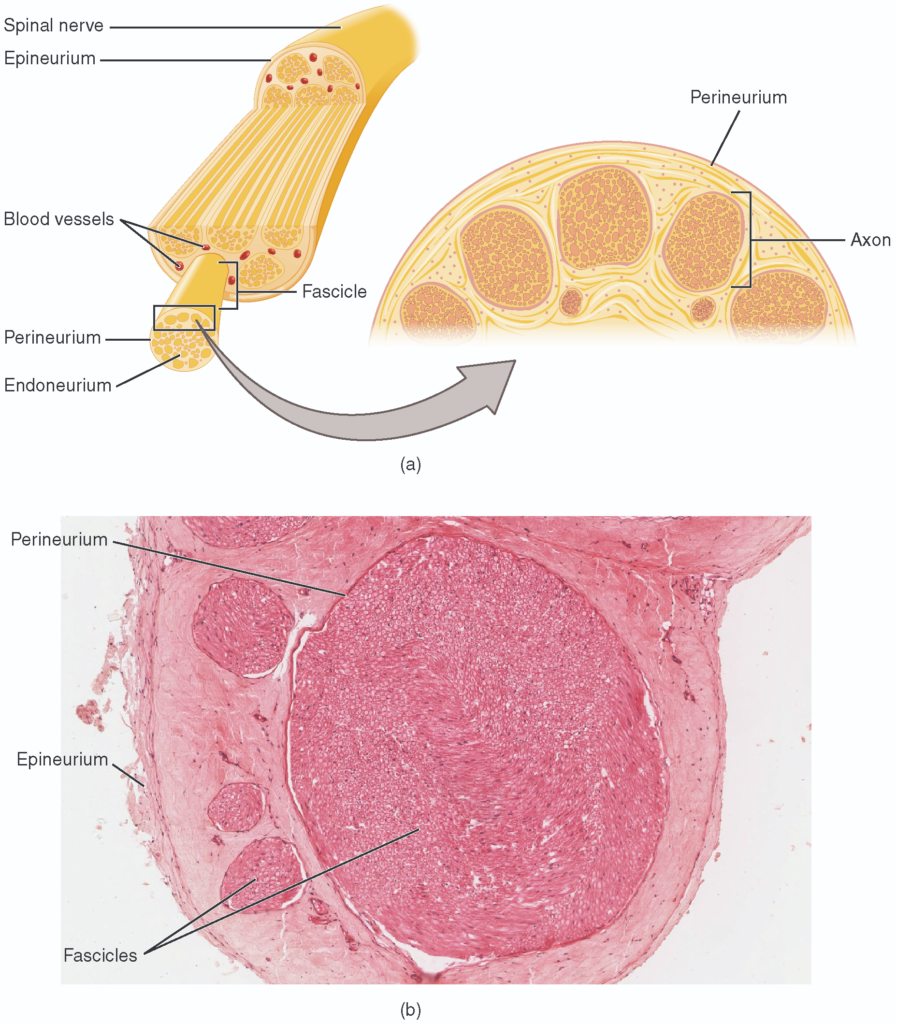
The structure of a nerve is organized by the layers of connective tissue on the outside, around each fascicle, and surrounding the individual nerve fibers (tissue source: simian). LM × 40. (Micrograph provided by the Regents of University of Michigan Medical School © 2012)

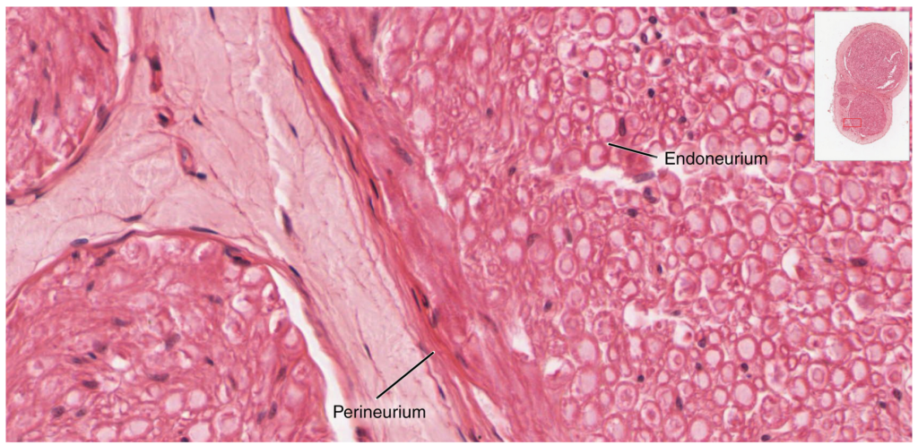
Zoom in on this slide of a nerve trunk to examine the endoneurium, perineurium, and epineurium in greater detail (tissue source: simian). LM × 1600. (Micrograph provided by the Regents of University of Michigan Medical School © 2012)

The anatomical arrangement of the roots of the cranial nerves observed from an inferior view of the brain.

Mnemonic#NameFunction (S/M/B)Central connection (nuclei)Peripheral connection (ganglion or muscle)
OnIOlfactorySmell (S)Olfactory bulbOlfactory epithelium
OldIIOpticVision (S)Hypothalamus, thalamus, midbrainRetina (retinal ganglion cells)
Olympus’IIIOculomotorEye movements (M)Oculomotor nucleusExtraocular muscles (other 4), levator palpebrae superioris, ciliary ganglion (autonomic)
ToweringIVTrochlearEye movements (M)Trochlear nucleusSuperior oblique muscle
TopsVTrigeminalSensory/motor – face (B)Trigeminal nuclei in the midbrain, pons, and medullaTrigeminal
AVIAbducensEye movements (M)Abducens nucleusLateral rectus muscle
FinnVIIFacialMotor – face, Taste (B)Facial nucleus, solitary nucleus, superior salivatory nucleusFacial muscles, Geniculate ganglion, Pterygopalatine ganglion (autonomic)
AndVIIIAuditory (Vestibulocochlear)Hearing/balance (S)Cochlear nucleus, Vestibular nucleus/cerebellumSpiral ganglion (hearing), Vestibular ganglion (balance)
GermanIXGlossopharyngealMotor – throat Taste (B)Solitary nucleus, inferior salivatory nucleus, nucleus ambiguusPharyngeal muscles, Geniculate ganglion, Otic ganglion (autonomic)
ViewedXVagusMotor/sensory – viscera (autonomic) (B)MedullaTerminal ganglia serving thoracic and upper abdominal organs (heart and small intestines)
SomeXISpinal AccessoryMotor – head and neck (M)Spinal accessory nucleusNeck muscles
HopsXIIHypoglossalMotor – lower throat (M)Hypoglossal nucleusMuscles of the larynx and lower pharynx

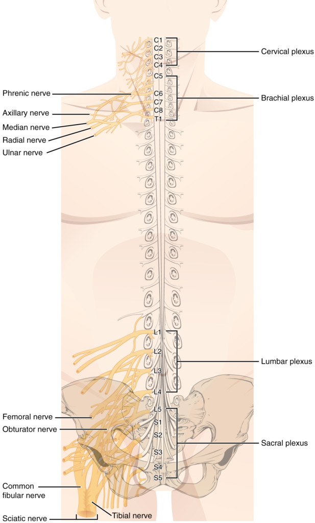
There are four main nerve plexuses in the human body. The cervical plexus supplies nerves to the posterior head and neck, as well as to the diaphragm. The brachial plexus supplies nerves to the arm. The lumbar plexus supplies nerves to the anterior leg. The sacral plexus supplies nerves to the posterior leg.

Nội dung này đang được cập nhật.
Dưới đây là video và các luyện tập (practice) của bài này. Nghe là một kĩ năng khó, đặc biệt là khi chúng ta chưa quen nội dung và chưa có nhạy cảm ngôn ngữ. Nhưng cứ đi thật chậm và đừng bỏ cuộc.
Xem video và cảm nhận nội dung bài. Bạn có thể thả trôi, cảm nhận dòng chảy ngôn ngữ và không nhất thiết phải hiểu toàn bộ bài. Bên dưới là script để bạn khái quát nội dụng và tra từ mới.
Script:
  1. The peripheral nervous system is composed of nerves and ganglia that are outside of the brain and spinal cord.
  2. Ganglia are groups of neurons and of two types, sensory or autonomic.
  3. Sensory ganglia contain unipolar sensory neurons and are found on the dorsal root of all spinal nerves as well as associated with many of the cranial nerves.
  4. Autonomic ganglia are in the sympathetic chain, the associated paravertebral or prevertebral ganglia, or in terminal ganglia near or within the organs controlled by the autonomic nervous system.
  5. Nerves are classified as cranial nerves or spinal nerves on the basis of their connection to the brain or spinal cord, respectively.
  6. The twelve cranial nerves can be strictly sensory in function, strictly motor in function, or a combination of the two functions.
  7. Sensory fibers are axons of sensory ganglia that carry sensory information into the brain and target sensory nuclei.
  8. Motor fibers are axons of motor neurons in motor nuclei of the brain stem and target skeletal muscles of the head and neck.
  9. Spinal nerves are all mixed nerves with both sensory and motor fibers.
  10. Spinal nerves emerge from the spinal cord and reorganize through plexuses, which then give rise to systemic nerves.
  11. Thoracic spinal nerves are not part of any plexus, but give rise to the intercostal nerves directly.
Bật video, nghe và điền từ vào chỗ trống.
Dưới đây là phần bàn luận. Bạn có thể tự do đặt câu hỏi, bổ sung kiến thức, và chia sẻ trải nghiệm của mình.
Subscribe
Notify of

0 Comments
Inline Feedbacks
View all comments

Ấn vào ô bên dưới để đánh dấu bạn đã hoàn thành bài học này

Quá dữ! Tiếp tục duy trì phong độ nhé!