Module 11: The Neurological Exam

Lesson 5: The Coordination and Gait Exams

Khám Phối Hợp Vận Động Và Dáng Đi

Nội dung bài học:
Mỗi bài học (lesson) bao gồm 4 phần chính: Thuật ngữ, Luyện Đọc, Luyện Nghe, và Bàn Luận.
Sử dụng tính năng:
Bôi hoặc nhấp đôi vào từ, sau đó ấn vào biểu tượng để tra nghĩa từ đó. Khi bạn đưa chuột đến câu (hoặc khi nhấp vào câu trên màn hình điện thoại), gợi ý về cách hiểu câu đó sẽ hiện lên. Cuối cùng, bạn có thể đánh dấu hoàn thành toàn bộ bài học bằng cách ấn vào nút “Hoàn Thành” ở cuối trang.
Đăng ký và đăng nhập
Bạn cần đăng ký và đăng nhập vào tài khoản để lưu quá trình học.
Dưới đây là danh sách những thuật ngữ Y khoa của module The Neurological Exam.
Khái quát được số lượng thuật ngữ sẽ xuất hiện trong bài đọc và nghe sẽ giúp bạn thoải mái tiêu thụ nội dung hơn. Sau khi hoàn thành nội dung đọc và nghe, bạn hãy quay lại đây và luyện tập (practice) để quen dần các thuật ngữ này. Đừng ép bản thân phải nhớ các thuật ngữ này vội vì bạn sẽ gặp và ôn lại danh sách này trong những bài học (lesson) khác của cùng một module.

Medical Terminology: The Neurological Exam

accommodation
in vision, a change in the ability of the eye to focus on objects at different distances
accommodation–convergence reflex
coordination of somatic control of the medial rectus muscles of either eye with the parasympathetic control of the ciliary bodies to maintain focus while the eyes converge on visual stimuli near to the face
anterograde amnesia
inability to form new memories from a particular time forward
aphasia
loss of language function
ataxia
movement disorder related to damage of the cerebellum characterized by loss of coordination in voluntary movements
Babinski sign
dorsiflexion of the foot with extension and splaying of the toes in response to the plantar reflex, normally suppressed by corticospinal input
cerebrocerebellum
lateral regions of the cerebellum; named for the significant input from the cerebral cortex
check reflex
response to a release in resistance so that the contractions stop, or check, movement
clasp-knife response
sign of UMN disease when a patient initially resists passive movement of a muscle but will quickly release to a lower state of resistance
conduction aphasia
loss of language function related to connecting the understanding of speech with the production of speech, without either specific function being lost
conductive hearing
hearing dependent on the conduction of vibrations of the tympanic membrane through the ossicles of the middle ear
conjugate gaze
coordinated movement of the two eyes simultaneously in the same direction
convergence
in vision, the movement of the eyes so that they are both pointed at the same point in space, which increases for stimuli that are closer to the subject
coordination exam
major section of the neurological exam that assesses complex, coordinated motor functions of the cerebellum and associated motor pathways
cortico-ponto-cerebellar pathway
projection from the cerebral cortex to the cerebellum by way of the gray matter of the pons
cranial nerve exam
major section of the neurological exam that assesses sensory and motor functions of the cranial nerves and their associated central and peripheral structures
cytoarchitecture
study of a tissue based on the structure and organization of its cellular components; related to the broader term, histology
deep tendon reflex
another term for stretch reflex, based on the elicitation through deep stimulation of the tendon at the insertion
diplopia
double vision resulting from a failure in conjugate gaze
edema
fluid accumulation in tissue; often associated with circulatory deficits
embolus
obstruction in a blood vessel such as a blood clot, fatty mass, air bubble, or other foreign matter that interrupts the flow of blood to an organ or some part of the body
episodic memory
memory of specific events in an autobiographical sense
expressive aphasia
loss of the ability to produce language; usually associated with damage to Broca’s area in the frontal lobe
extrinsic muscles of the tongue
muscles that are connected to other structures, such as the hyoid bone or the mandible, and control the position of the tongue
fasciculation
small muscle twitch as a result of spontaneous activity from an LMN
fauces
opening from the oral cavity into the pharynx
fibrillation
in motor responses, a spontaneous muscle action potential that occurs in the absence of neuromuscular input, resulting from LMN lesions
flaccid paralysis
loss of voluntary muscle control and muscle tone, as the result of LMN disease
flaccidity
presentation of a loss of muscle tone, observed as floppy limbs or a lack of resistance to passive movement
flocculonodular lobe
lobe of the cerebellum that receives input from the vestibular system to help with balance and posture
gait
rhythmic pattern of alternating movements of the lower limbs during locomotion
gait exam
major section of the neurological exam that assesses the cerebellum and descending pathways in the spinal cord through the coordinated motor functions of walking; a portion of the coordination exam
gnosis
in a neurological exam, intuitive experiential knowledge tested by interacting with common objects or symbols
graphesthesia
perception of symbols, such as letters or numbers, traced in the palm of the hand
hemisection
cut through half of a structure, such as the spinal cord
hemorrhagic stroke
disruption of blood flow to the brain caused by bleeding within the cranial vault
hyperflexia
overly flexed joints
hypotonicity
low muscle tone, a sign of LMN disease
hypovolemia
decrease in blood volume
inferior cerebellar peduncle (ICP)
input to the cerebellum, largely from the inferior olive, that represents sensory feedback from the periphery
inferior olive
large nucleus in the medulla that receives input from sensory systems and projects into the cerebellar cortex
internuclear ophthalmoplegia
deficit of conjugate lateral gaze because the lateral rectus muscle of one eye does not contract resulting from damage to the abducens nerve or the MLF
intorsion
medial rotation of the eye around its axis
intrinsic muscles of the tongue
muscles that originate out of, and insert into, other tissues within the tongue and control the shape of the tongue
ischemic stroke
disruption of blood flow to the brain because blood cannot flow through blood vessels as a result of a blockage or narrowing of the vessel
jaw-jerk reflex
stretch reflex of the masseter muscle
localization of function
principle that circumscribed anatomical locations are responsible for specific functions in an organ system
medial longitudinal fasciculus (MLF)
fiber pathway that connects structures involved in the control of eye and head position, from the superior colliculus to the vestibular nuclei and cerebellum
mental status exam
major section of the neurological exam that assesses cognitive functions of the cerebrum
middle cerebellar peduncle (MCP)
large, white-matter bridge from the pons that constitutes the major input to the cerebellar cortex
motor exam
major section of the neurological exam that assesses motor functions of the spinal cord and spinal nerves
neurological exam
clinical assessment tool that can be used to quickly evaluate neurological function and determine if specific parts of the nervous system have been affected by damage or disease
paramedian pontine reticular formation (PPRF)
region of the brain stem adjacent to the motor nuclei for gaze control that coordinates rapid, conjugate eye movements
paresis
partial loss of, or impaired, voluntary muscle control
plantar reflex
superficial reflex initiated by gentle stimulation of the sole of the foot
praxis
in a neurological exam, the act of doing something using ready knowledge or skills in response to verbal instruction
procedural memory
memory of how to perform a specific task
pronator drift
sign of contralateral corticospinal lesion when the one arm will drift into a pronated position when held straight out with the palms facing upward
receptive aphasia
loss of the ability to understand received language, such as what is spoken to the subject or given in written form
red nucleus
nucleus in the midbrain that receives output from the cerebellum and projects onto the spinal cord in the rubrospinal tract
retrograde amnesia
loss of memories before a particular event
Rinne test
use of a tuning fork to test conductive hearing loss versus sensorineural hearing loss
Romberg test
test of equilibrium that requires the patient to maintain a straight, upright posture without visual feedback of position
rubrospinal tract
descending tract from the red nucleus of the midbrain that results in modification of ongoing motor programs
saccade
small, rapid movement of the eyes used to locate and direct the fovea onto visual stimuli
sensorineural hearing
hearing dependent on the transduction and propagation of auditory information through the neural components of the peripheral auditory structures
sensory exam
major section of the neurological exam that assesses sensory functions of the spinal cord and spinal nerves
short-term memory
capacity to retain information actively in the brain for a brief period of time
Snellen chart
standardized arrangement of letters in decreasing size presented to a subject at a distance of 20 feet to test visual acuity
spasticity
increased contraction of a muscle in response to resistance, often resulting in hyperflexia
spinocerebellar tract
ascending fibers that carry proprioceptive input to the cerebellum used in maintaining balance and coordinated movement
spinocerebellum
midline region of the cerebellum known as the vermis that receives proprioceptive input from the spinal cord
stereognosis
perception of common objects placed in the hand solely on the basis of manipulation of that object in the hand
stroke
(also, cerebrovascular accident (CVA)) loss of neurological function caused by an interruption of blood flow to a region of the central nervous system
superficial reflex
reflexive contraction initiated by gentle stimulation of the skin
superior cerebellar peduncle (SCP)
white-matter tract representing output of the cerebellum to the red nucleus of the midbrain
transient ischemic attack (TIA)
temporary disruption of blood flow to the brain in which symptoms occur rapidly but last only a short time
vermis
prominent ridge along the midline of the cerebellum that is referred to as the spinocerebellum
vestibulo-ocular reflex (VOR)
reflex based on connections between the vestibular system and the cranial nerves of eye movements that ensures that images are stabilized on the retina as the head and body move
vestibulocerebellum
flocculonodular lobe of the cerebellum named for the vestibular input from the eighth cranial nerve
Weber test
use of a tuning fork to test the laterality of hearing loss by placing it at several locations on the midline of the skull
Wernicke’s area
region at the posterior end of the lateral sulcus in which speech comprehension is localized
Nội dung này đang được cập nhật.
Dưới đây là các bài văn nằm ở bên trái. Ở bên phải là các bài luyện tập (practice) để đánh giá khả năng đọc hiểu của bạn. Sẽ khó khăn trong thời gian đầu nếu vốn từ vựng của bạn còn hạn chế, đặc biệt là từ vựng Y khoa. Hãy kiên nhẫn và đọc nhiều nhất có kể, lượng kiến thức tích tụ dần sẽ giúp bạn đọc thoải mái hơn.
The role of the cerebellum is a subject of debate. There is an obvious connection to motor function based on the clinical implications of cerebellar damage. There is also strong evidence of the cerebellar role in procedural memory. The two are not incompatible; in fact, procedural memory is motor memory, such as learning to ride a bicycle. Significant work has been performed to describe the connections within the cerebellum that result in learning. A model for this learning is classical conditioning, as shown by the famous dogs from the physiologist Ivan Pavlov’s work. This classical conditioning, which can be related to motor learning, fits with the neural connections of the cerebellum. The cerebellum is 10 percent of the mass of the brain and has varied functions that all point to a role in the motor system.
The cerebellum is located in apposition to the dorsal surface of the brain stem, centered on the pons. The name of the pons is derived from its connection to the cerebellum. The word means “bridge” and refers to the thick bundle of myelinated axons that form a bulge on its ventral surface. Those fibers are axons that project from the gray matter of the pons into the contralateral cerebellar cortex. These fibers make up the middle cerebellar peduncle (MCP) and are the major physical connection of the cerebellum to the brain stem (Figure 1). Two other white matter bundles connect the cerebellum to the other regions of the brain stem. The superior cerebellar peduncle (SCP) is the connection of the cerebellum to the midbrain and forebrain. The inferior cerebellar peduncle (ICP) is the connection to the medulla.

These connections can also be broadly described by their functions. The ICP conveys sensory input to the cerebellum, partially from the spinocerebellar tract, but also through fibers of the inferior olive. The MCP is part of the cortico-ponto-cerebellar pathway that connects the cerebral cortex with the cerebellum and preferentially targets the lateral regions of the cerebellum. It includes a copy of the motor commands sent from the precentral gyrus through the corticospinal tract, arising from collateral branches that synapse in the gray matter of the pons, along with input from other regions such as the visual cortex. The SCP is the major output of the cerebellum, divided between the red nucleus in the midbrain and the thalamus, which will return cerebellar processing to the motor cortex. These connections describe a circuit that compares motor commands and sensory feedback to generate a new output. These comparisons make it possible to coordinate movements. If the cerebral cortex sends a motor command to initiate walking, that command is copied by the pons and sent into the cerebellum through the MCP. Sensory feedback in the form of proprioception from the spinal cord, as well as vestibular sensations from the inner ear, enters through the ICP. If you take a step and begin to slip on the floor because it is wet, the output from the cerebellum—through the SCP—can correct for that and keep you balanced and moving. The red nucleus sends new motor commands to the spinal cord through the rubrospinal tract.

The cerebellum is divided into regions that are based on the particular functions and connections involved. The midline regions of the cerebellum, the vermis and flocculonodular lobe, are involved in comparing visual information, equilibrium, and proprioceptive feedback to maintain balance and coordinate movements such as walking, or gait, through the descending output of the red nucleus (Figure 2). The lateral hemispheres are primarily concerned with planning motor functions through frontal lobe inputs that are returned through the thalamic projections back to the premotor and motor cortices. Processing in the midline regions targets movements of the axial musculature, whereas the lateral regions target movements of the appendicular musculature. The vermis is referred to as the spinocerebellum because it primarily receives input from the dorsal columns and spinocerebellar pathways. The flocculonodular lobe is referred to as the vestibulocerebellum because of the vestibular projection into that region. Finally, the lateral cerebellum is referred to as the cerebrocerebellum, reflecting the significant input from the cerebral cortex through the cortico-ponto-cerebellar pathway.
Testing for cerebellar function is the basis of the coordination exam. The subtests target appendicular musculature, controlling the limbs, and axial musculature for posture and gait. The assessment of cerebellar function will depend on the normal functioning of other systems addressed in previous sections of the neurological exam. Motor control from the cerebrum, as well as sensory input from somatic, visual, and vestibular senses, are important to cerebellar function.

The subtests that address appendicular musculature, and therefore the lateral regions of the cerebellum, begin with a check for tremor. The patient extends their arms in front of them and holds the position. The examiner watches for the presence of tremors that would not be present if the muscles are relaxed. By pushing down on the arms in this position, the examiner can check for the rebound response, which is when the arms are automatically brought back to the extended position. The extension of the arms is an ongoing motor process, and the tap or push on the arms presents a change in the proprioceptive feedback. The cerebellum compares the cerebral motor command with the proprioceptive feedback and adjusts the descending input to correct. The red nucleus would send an additional signal to the LMN for the arm to increase contraction momentarily to overcome the change and regain the original position.

The check reflex depends on cerebellar input to keep increased contraction from continuing after the removal of resistance. The patient flexes the elbow against resistance from the examiner to extend the elbow. When the examiner releases the arm, the patient should be able to stop the increased contraction and keep the arm from moving. A similar response would be seen if you try to pick up a coffee mug that you believe to be full but turns out to be empty. Without checking the contraction, the mug would be thrown from the overexertion of the muscles expecting to lift a heavier object.

Several subtests of the cerebellum assess the ability to alternate movements, or switch between muscle groups that may be antagonistic to each other. In the finger-to-nose test, the patient touches their finger to the examiner’s finger and then to their nose, and then back to the examiner’s finger, and back to the nose. The examiner moves the target finger to assess a range of movements. A similar test for the lower extremities has the patient touch their toe to a moving target, such as the examiner’s finger. Both of these tests involve flexion and extension around a joint—the elbow or the knee and the shoulder or hip—as well as movements of the wrist and ankle. The patient must switch between the opposing muscles, like the biceps and triceps brachii, to move their finger from the target to their nose. Coordinating these movements involves the motor cortex communicating with the cerebellum through the pons and feedback through the thalamus to plan the movements. Visual cortex information is also part of the processing that occurs in the cerebrocerebellum while it is involved in guiding movements of the finger or toe.

Rapid, alternating movements are tested for the upper and lower extremities. The patient is asked to touch each finger to their thumb, or to pat the palm of one hand on the back of the other, and then flip that hand over and alternate back-and-forth. To test similar function in the lower extremities, the patient touches their heel to their shin near the knee and slides it down toward the ankle, and then back again, repetitively. Rapid, alternating movements are part of speech as well. A patient is asked to repeat the nonsense consonants “lah-kah-pah” to alternate movements of the tongue, lips, and palate. All of these rapid alternations require planning from the cerebrocerebellum to coordinate movement commands that control the coordination.
Gait can either be considered a separate part of the neurological exam or a subtest of the coordination exam that addresses walking and balance. Testing posture and gait addresses functions of the spinocerebellum and the vestibulocerebellum because both are part of these activities. A subtest called station begins with the patient standing in a normal position to check for the placement of the feet and balance. The patient is asked to hop on one foot to assess the ability to maintain balance and posture during movement. Though the station subtest appears to be similar to the Romberg test, the difference is that the patient’s eyes are open during station. The Romberg test has the patient stand still with the eyes closed. Any changes in posture would be the result of proprioceptive deficits, and the patient is able to recover when they open their eyes.

Subtests of walking begin with having the patient walk normally for a distance away from the examiner, and then turn and return to the starting position. The examiner watches for abnormal placement of the feet and the movement of the arms relative to the movement. The patient is then asked to walk with a few different variations. Tandem gait is when the patient places the heel of one foot against the toe of the other foot and walks in a straight line in that manner. Walking only on the heels or only on the toes will test additional aspects of balance.
A movement disorder of the cerebellum is referred to as ataxia. It presents as a loss of coordination in voluntary movements. Ataxia can also refer to sensory deficits that cause balance problems, primarily in proprioception and equilibrium. When the problem is observed in movement, it is ascribed to cerebellar damage. Sensory and vestibular ataxia would likely also present with problems in gait and station.

Ataxia is often the result of exposure to exogenous substances, focal lesions, or a genetic disorder. Focal lesions include strokes affecting the cerebellar arteries, tumors that may impinge on the cerebellum, trauma to the back of the head and neck, or MS. Alcohol intoxication or drugs such as ketamine cause ataxia, but it is often reversible. Mercury in fish can cause ataxia as well. Hereditary conditions can lead to degeneration of the cerebellum or spinal cord, as well as malformation of the brain, or the abnormal accumulation of copper seen in Wilson’s disease.

OpenStax. (2022). Anatomy and Physiology 2e. Rice University. Retrieved June 15, 2023. ISBN-13: 978-1-711494-06-7 (Hardcover) ISBN-13: 978-1-711494-05-0 (Paperback) ISBN-13: 978-1-951693-42-8 (Digital). License: Attribution 4.0 International (CC BY 4.0). Access for free at openstax.org.

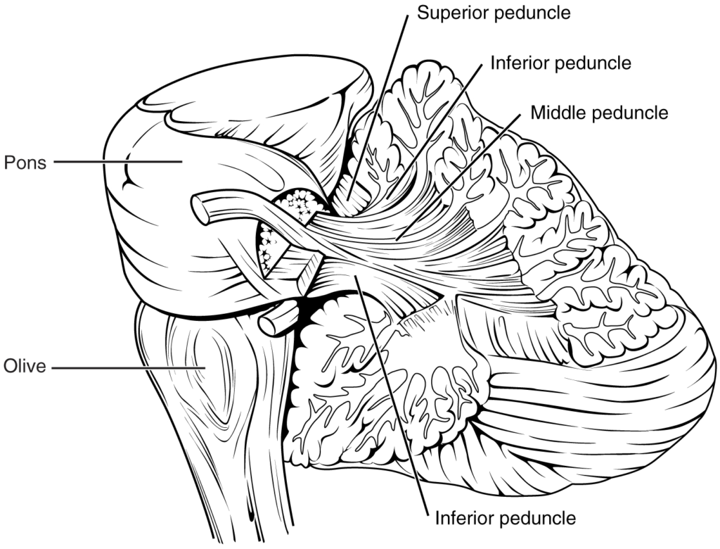
The connections to the cerebellum are the three cerebellar peduncles, which are close to each other. The ICP arises from the medulla—specifically from the inferior olive, which is visible as a bulge on the ventral surface of the brain stem. The MCP is the ventral surface of the pons. The SCP projects into the midbrain.

The cerebellum can be divided into two basic regions: the midline and the hemispheres. The midline is composed of the vermis and the flocculonodular lobe, and the hemispheres are the lateral regions.

Nội dung này đang được cập nhật.
Dưới đây là video và các luyện tập (practice) của bài này. Nghe là một kĩ năng khó, đặc biệt là khi chúng ta chưa quen nội dung và chưa có nhạy cảm ngôn ngữ. Nhưng cứ đi thật chậm và đừng bỏ cuộc.
Xem video và cảm nhận nội dung bài. Bạn có thể thả trôi, cảm nhận dòng chảy ngôn ngữ và không nhất thiết phải hiểu toàn bộ bài. Bên dưới là script để bạn khái quát nội dụng và tra từ mới.
Script:
  1. The cerebellum is an important part of motor function in the nervous system.
  2. It apparently plays a role in procedural learning, which would include motor skills such as riding a bike or throwing a football.
  3. The basis for these roles is likely to be tied into the role the cerebellum plays as a comparator for voluntary movement.
  4. The motor commands from the cerebral hemispheres travel along the corticospinal pathway, which passes through the pons.
  5. Collateral branches of these fibers synapse on neurons in the pons, which then project into the cerebellar cortex through the middle cerebellar peduncles.
  6. Ascending sensory feedback, entering through the inferior cerebellar peduncles, provides information about motor performance.
  7. The cerebellar cortex compares the command to the actual performance and can adjust the descending input to compensate for any mismatch.
  8. The output from deep cerebellar nuclei projects through the superior cerebellar peduncles to initiate descending signals from the red nucleus to the spinal cord.
  9. The primary role of the cerebellum in relation to the spinal cord is through the spinocerebellum.
  10. It controls posture and gait with significant input from the vestibular system.
  11. Deficits in cerebellar function result in ataxias, or a specific kind of movement disorder.
  12. The root cause of the ataxia may be the sensory input.
  13. This could be either the proprioceptive input from the spinal cord or the equilibrium input from the vestibular system, or direct damage to the cerebellum by stroke, trauma, hereditary factors, or toxins.
Bật video, nghe và điền từ vào chỗ trống.
Dưới đây là phần bàn luận. Bạn có thể tự do đặt câu hỏi, bổ sung kiến thức, và chia sẻ trải nghiệm của mình.
Subscribe
Notify of

0 Comments
Inline Feedbacks
View all comments

Ấn vào ô bên dưới để đánh dấu bạn đã hoàn thành bài học này

Quá dữ! Tiếp tục duy trì phong độ nhé!