Module 16: The Urinary System

Lesson 4: Microscopic Anatomy of the Kidney

Giải Phẫu Vi Thể Thận

Nội dung bài học:
Mỗi bài học (lesson) bao gồm 4 phần chính: Thuật ngữ, Luyện Đọc, Luyện Nghe, và Bàn Luận.
Sử dụng tính năng:
Bôi hoặc nhấp đôi vào từ, sau đó ấn vào biểu tượng để tra nghĩa từ đó. Khi bạn đưa chuột đến câu (hoặc khi nhấp vào câu trên màn hình điện thoại), gợi ý về cách hiểu câu đó sẽ hiện lên. Cuối cùng, bạn có thể đánh dấu hoàn thành toàn bộ bài học bằng cách ấn vào nút “Hoàn Thành” ở cuối trang.
Đăng ký và đăng nhập
Bạn cần đăng ký và đăng nhập vào tài khoản để lưu quá trình học.
Dưới đây là danh sách những thuật ngữ Y khoa của module The Urinary System.
Khái quát được số lượng thuật ngữ sẽ xuất hiện trong bài đọc và nghe sẽ giúp bạn thoải mái tiêu thụ nội dung hơn. Sau khi hoàn thành nội dung đọc và nghe, bạn hãy quay lại đây và luyện tập (practice) để quen dần các thuật ngữ này. Đừng ép bản thân phải nhớ các thuật ngữ này vội vì bạn sẽ gặp và ôn lại danh sách này trong những bài học (lesson) khác của cùng một module.

Medical Terminology: The Urinary System

anatomical sphincter
smooth or skeletal muscle surrounding the lumen of a vessel or hollow organ that can restrict flow when contracted
angiotensin I
protein produced by the enzymatic action of renin on angiotensinogen; inactive precursor of angiotensin II
angiotensin II
protein produced by the enzymatic action of ACE on inactive angiotensin I; actively causes vasoconstriction and stimulates aldosterone release by the adrenal cortex
angiotensin-converting enzyme (ACE)
enzyme produced by the lungs that catalyzes the reaction of inactive angiotensin I into active angiotensin II
angiotensinogen
inactive protein in the circulation produced by the liver; precursor of angiotensin I; must be modified by the enzymes renin and ACE to be activated
anuria
absence of urine produced; production of 50 mL or less per day
aquaporin
protein-forming water channels through the lipid bilayer of the cell; allows water to cross; activation in the collecting ducts is under the control of ADH
Bowman’s capsule
cup-shaped sack lined by a simple squamous epithelium (parietal surface) and specialized cells called podocytes (visceral surface) that participate in the filtration process; receives the filtrate which then passes on to the PCTs
brush border
formed by microvilli on the surface of certain cuboidal cells; in the kidney it is found in the PCT; increases surface area for absorption in the kidney
calyces
cup-like structures receiving urine from the collecting ducts where it passes on to the renal pelvis and ureter
cortical nephrons
nephrons with loops of Henle that do not extend into the renal medulla
countercurrent multiplier system
involves the descending and ascending loops of Henle directing forming urine in opposing directions to create a concentration gradient when combined with variable permeability and sodium pumping
detrusor muscle
smooth muscle in the bladder wall; fibers run in all directions to reduce the size of the organ when emptying it of urine
distal convoluted tubules
portions of the nephron distal to the loop of Henle that receive hyposmotic filtrate from the loop of Henle and empty into collecting ducts
diuretic
compound that increases urine output, leading to decreased water conservation
efferent arteriole
arteriole carrying blood from the glomerulus to the capillary beds around the convoluted tubules and loop of Henle; portion of the portal system
endothelins
group of vasoconstrictive, 21-amino acid peptides; produced by endothelial cells of the renal blood vessels, mesangial cells, and cells of the DCT
external urinary sphincter
skeletal muscle; must be relaxed consciously to void urine
fenestrations
small windows through a cell, allowing rapid filtration based on size; formed in such a way as to allow substances to cross through a cell without mixing with cell contents
filtration slits
formed by pedicels of podocytes; substances filter between the pedicels based on size
forming urine
filtrate undergoing modifications through secretion and reabsorption before true urine is produced
glomerular filtration rate (GFR)
rate of renal filtration
glomerulus
tuft of capillaries surrounded by Bowman’s capsule; filters the blood based on size
glycosuria
presence of glucose in the urine; caused by high blood glucose levels that exceed the ability of the kidneys to reabsorb the glucose; usually the result of untreated or poorly controlled diabetes mellitus
incontinence
loss of ability to control micturition
intercalated cell
specialized cell of the collecting ducts that secrete or absorb acid or bicarbonate; important in acid–base balance
internal urinary sphincter
smooth muscle at the juncture of the bladder and urethra; relaxes as the bladder fills to allow urine into the urethra
inulin
plant polysaccharide injected to determine GFR; is neither secreted nor absorbed by the kidney, so its appearance in the urine is directly proportional to its filtration rate
juxtaglomerular apparatus (JGA)
located at the juncture of the DCT and the afferent and efferent arterioles of the glomerulus; plays a role in the regulation of renal blood flow and GFR
juxtaglomerular cell
modified smooth muscle cells of the afferent arteriole; secretes renin in response to a drop in blood pressure
juxtamedullary nephrons
nephrons adjacent to the border of the cortex and medulla with loops of Henle that extend into the renal medulla
leaky tight junctions
tight junctions in which the sealing strands of proteins between the membranes of adjacent cells are fewer in number and incomplete; allows limited intercellular movement of solvent and solutes
leukocyte esterase
enzyme produced by leukocytes that can be detected in the urine and that serves as an indirect indicator of urinary tract infection
loop of Henle
descending and ascending portions between the proximal and distal convoluted tubules; those of cortical nephrons do not extend into the medulla, whereas those of juxtamedullary nephrons do extend into the medulla
macula densa
cells found in the part of the DCT forming the JGA; sense Na+ concentration in the forming urine
medulla
inner region of kidney containing the renal pyramids
mesangial
contractile cells found in the glomerulus; can contract or relax to regulate filtration rate
micturition
also called urination or voiding
myogenic mechanism
mechanism by which smooth muscle responds to stretch by contracting; an increase in blood pressure causes vasoconstriction and a decrease in blood pressure causes vasodilation so that blood flow downstream remains steady
nephrons
functional units of the kidney that carry out all filtration and modification to produce urine; consist of renal corpuscles, proximal and distal convoluted tubules, and descending and ascending loops of Henle; drain into collecting ducts
net filtration pressure (NFP)
pressure of fluid across the glomerulus; calculated by taking the hydrostatic pressure of the capillary and subtracting the colloid osmotic pressure of the blood and the hydrostatic pressure of Bowman’s capsule
oliguria
below normal urine production of 400–500 mL/day
osteomalacia
softening of bones due to a lack of mineralization with calcium and phosphate; most often due to lack of vitamin D; in children, osteomalacia is termed rickets; not to be confused with osteoporosis
pedicels
finger-like projections of podocytes surrounding glomerular capillaries; interdigitate to form a filtration membrane
peritubular capillaries
second capillary bed of the renal portal system; surround the proximal and distal convoluted tubules; associated with the vasa recta
physiological sphincter
sphincter consisting of circular smooth muscle indistinguishable from adjacent muscle but possessing differential innervations, permitting its function as a sphincter; structurally weak
podocytes
cells forming finger-like processes; form the visceral layer of Bowman’s capsule; pedicels of the podocytes interdigitate to form a filtration membrane
polyuria
urine production in excess of 2.5 L/day; may be caused by diabetes insipidus, diabetes mellitus, or excessive use of diuretics
principal cell
found in collecting ducts and possess channels for the recovery or loss of sodium and potassium; under the control of aldosterone; also have aquaporin channels under ADH control to regulate recovery of water
proximal convoluted tubules (PCTs)
tortuous tubules receiving filtrate from Bowman’s capsule; most active part of the nephron in reabsorption and secretion
renal columns
extensions of the renal cortex into the renal medulla; separates the renal pyramids; contains blood vessels and connective tissues
renal corpuscle
consists of the glomerulus and Bowman’s capsule
renal cortex
outer part of kidney containing all of the nephrons; some nephrons have loops of Henle extending into the medulla
renal fat pad
adipose tissue between the renal fascia and the renal capsule that provides protective cushioning to the kidney
renal hilum
recessed medial area of the kidney through which the renal artery, renal vein, ureters, lymphatics, and nerves pass
renal papillae
medullary area of the renal pyramids where collecting ducts empty urine into the minor calyces
renal pyramids
six to eight cone-shaped tissues in the medulla of the kidney containing collecting ducts and the loops of Henle of juxtamedullary nephrons
renin
enzyme produced by juxtaglomerular cells in response to decreased blood pressure or sympathetic nervous activity; catalyzes the conversion of angiotensinogen into angiotensin I
retroperitoneal
behind the peritoneum; in the case of the kidney and ureters, between the parietal peritoneum and the abdominal wall
sacral micturition center
group of neurons in the sacral region of the spinal cord that controls urination; acts reflexively unless its action is modified by higher brain centers to allow voluntary urination
specific gravity
weight of a liquid compared to pure water, which has a specific gravity of 1.0; any solute added to water will increase its specific gravity
systemic edema
increased fluid retention in the interstitial spaces and cells of the body; can be seen as swelling over large areas of the body, particularly the lower extremities
trigone
area at the base of the bladder marked by the two ureters in the posterior–lateral aspect and the urethral orifice in the anterior aspect oriented like points on a triangle
tubuloglomerular feedback
feedback mechanism involving the JGA; macula densa cells monitor Na+ concentration in the terminal portion of the ascending loop of Henle and act to cause vasoconstriction or vasodilation of afferent and efferent arterioles to alter GFR
urethra
transports urine from the bladder to the outside environment
urinalysis
analysis of urine to diagnose disease
urochrome
heme-derived pigment that imparts the typical yellow color of urine
vasa recta
branches of the efferent arterioles that parallel the course of the loops of Henle and are continuous with the peritubular capillaries; with the glomerulus, form a portal system
Nội dung này đang được cập nhật.
Dưới đây là các bài văn nằm ở bên trái. Ở bên phải là các bài luyện tập (practice) để đánh giá khả năng đọc hiểu của bạn. Sẽ khó khăn trong thời gian đầu nếu vốn từ vựng của bạn còn hạn chế, đặc biệt là từ vựng Y khoa. Hãy kiên nhẫn và đọc nhiều nhất có kể, lượng kiến thức tích tụ dần sẽ giúp bạn đọc thoải mái hơn.
The renal structures that conduct the essential work of the kidney cannot be seen by the naked eye. Only a light or electron microscope can reveal these structures. Even then, serial sections and computer reconstruction are necessary to give us a comprehensive view of the functional anatomy of the nephron and its associated blood vessels.

Nephrons are the functional units of the kidneys. They take a simple filtrate of the blood and modify it into urine. Many changes take place in the different parts of the nephron before urine is created for disposal. The term forming urine will be used hereafter to describe the filtrate as it is modified into true urine. The principle task of the nephron population is to balance the plasma to homeostatic set points and excrete potential toxins in the urine. They do this by accomplishing three principle functions—filtration, reabsorption, and secretion. They also have additional secondary functions that exert control in three areas: blood pressure (via production of renin), red blood cell production (via the hormone EPO), and calcium absorption (via conversion of calcidiol into calcitriol, the active form of vitamin D).
As discussed earlier, the renal corpuscle consists of a tuft of capillaries called the glomerulus that is largely surrounded by Bowman’s (glomerular) capsule. The glomerulus is a high-pressure capillary bed between afferent and efferent arterioles. Bowman’s capsule surrounds the glomerulus to form a lumen, and captures and directs this filtrate to the PCT. The outermost part of Bowman’s capsule, the parietal layer, is a simple squamous epithelium. It transitions onto the glomerular capillaries in an intimate embrace to form the visceral layer of the capsule. Here, the cells are not squamous, but uniquely shaped cells (podocytes) extending finger-like arms (pedicels) to cover the glomerular capillaries (Figure 1). These projections interdigitate to form filtration slits, leaving small gaps between the digits to form a sieve. As blood passes through the glomerulus, 10 to 20 percent of the plasma filters between these sieve-like fingers to be captured by Bowman’s capsule and funneled to the PCT. Where the fenestrae (windows) in the glomerular capillaries match the spaces between the podocyte “fingers,” the only thing separating the capillary lumen and the lumen of Bowman’s capsule is their shared basement membrane (Figure 2). These three features comprise what is known as the filtration membrane. This membrane permits very rapid movement of filtrate from capillary to capsule though pores that are only 70 nm in diameter.

The fenestrations prevent filtration of blood cells or large proteins, but allow most other constituents through. These substances cross readily if they are less than 4 nm in size and most pass freely up to 8 nm in size. An additional factor affecting the ability of substances to cross this barrier is their electric charge. The proteins associated with these pores are negatively charged, so they tend to repel negatively charged substances and allow positively charged substances to pass more readily. The basement membrane prevents filtration of medium-to-large proteins such as globulins. There are also mesangial cells in the filtration membrane that can contract to help regulate the rate of filtration of the glomerulus. Overall, filtration is regulated by fenestrations in capillary endothelial cells, podocytes with filtration slits, membrane charge, and the basement membrane between capillary cells. The result is the creation of a filtrate that does not contain cells or large proteins, and has a slight predominance of positively charged substances.

Lying just outside Bowman’s capsule and the glomerulus is the juxtaglomerular apparatus (JGA) (Figure 3). At the juncture where the afferent and efferent arterioles enter and leave Bowman’s capsule, the initial part of the distal convoluted tubule (DCT) comes into direct contact with the arterioles. The wall of the DCT at that point forms a part of the JGA known as the macula densa. This cluster of cuboidal epithelial cells monitors the fluid composition of fluid flowing through the DCT. In response to the concentration of Na+ in the fluid flowing past them, these cells release paracrine signals. They also have a single, nonmotile cilium that responds to the rate of fluid movement in the tubule. The paracrine signals released in response to changes in flow rate and Na+ concentration are adenosine triphosphate (ATP) and adenosine.

A second cell type in this apparatus is the juxtaglomerular cell. This is a modified, smooth muscle cell lining the afferent arteriole that can contract or relax in response to ATP or adenosine released by the macula densa. Such contraction and relaxation regulate blood flow to the glomerulus. If the osmolarity of the filtrate is too high (hyperosmotic), the juxtaglomerular cells will contract, decreasing the glomerular filtration rate (GFR) so less plasma is filtered, leading to less urine formation and greater retention of fluid. This will ultimately decrease blood osmolarity toward the physiologic norm. If the osmolarity of the filtrate is too low, the juxtaglomerular cells will relax, increasing the GFR and enhancing the loss of water to the urine, causing blood osmolarity to rise. In other words, when blood osmolarity goes up, filtration and urine formation decrease and water is retained. When blood osmolarity goes down, filtration and urine formation increase and water is lost by way of the urine. The net result of these opposing actions is to keep the rate of filtration relatively constant. A second function of the macula densa cells is to regulate renin release from the juxtaglomerular cells of the afferent arteriole (Figure 4). Active renin is a protein comprised of 304 amino acids that cleaves several amino acids from angiotensinogen to produce angiotensin I. Angiotensin I is not biologically active until converted to angiotensin II by angiotensin-converting enzyme (ACE) from the lungs. Angiotensin II is a systemic vasoconstrictor that helps to regulate blood pressure by increasing it. Angiotensin II also stimulates the release of the steroid hormone aldosterone from the adrenal cortex. Aldosterone stimulates Na+ reabsorption by the kidney, which also results in water retention and increased blood pressure.
Filtered fluid collected by Bowman’s capsule enters into the PCT. It is called convoluted due to its tortuous path. Simple cuboidal cells form this tubule with prominent microvilli on the luminal surface, forming a brush border. These microvilli create a large surface area to maximize the absorption and secretion of solutes (Na+, Cl–, glucose, etc.), the most essential function of this portion of the nephron. These cells actively transport ions across their membranes, so they possess a high concentration of mitochondria in order to produce sufficient ATP.
The descending and ascending portions of the loop of Henle (sometimes referred to as the nephron loop) are, of course, just continuations of the same tubule. They run adjacent and parallel to each other after having made a hairpin turn at the deepest point of their descent. The descending loop of Henle consists of an initial short, thick portion and long, thin portion, whereas the ascending loop consists of an initial short, thin portion followed by a long, thick portion. The descending thick portion consists of simple cuboidal epithelium similar to that of the PCT. The descending and ascending thin portions consists of simple squamous epithelium. As you will see later, these are important differences, since different portions of the loop have different permeabilities for solutes and water. The ascending thick portion consists of simple cuboidal epithelium similar to the DCT.
The DCT, like the PCT, is very tortuous and formed by simple cuboidal epithelium, but it is shorter than the PCT. These cells are not as active as those in the PCT; thus, there are fewer microvilli on the apical surface. However, these cells must also pump ions against their concentration gradient, so you will find of large numbers of mitochondria, although fewer than in the PCT.
The collecting ducts are continuous with the nephron but not technically part of it. In fact, each duct collects filtrate from several nephrons for final modification. Collecting ducts merge as they descend deeper in the medulla to form about 30 terminal ducts, which empty at a papilla. They are lined with simple squamous epithelium with receptors for ADH. When stimulated by ADH, these cells will insert aquaporin channel proteins into their membranes, which as their name suggests, allow water to pass from the duct lumen through the cells and into the interstitial spaces to be recovered by the vasa recta. This process allows for the recovery of large amounts of water from the filtrate back into the blood. In the absence of ADH, these channels are not inserted, resulting in the excretion of water in the form of dilute urine. Most, if not all, cells of the body contain aquaporin molecules, whose channels are so small that only water can pass. At least 10 types of aquaporins are known in humans, and six of those are found in the kidney. The function of all aquaporins is to allow the movement of water across the lipid-rich, hydrophobic cell membrane (Figure 5).

OpenStax. (2022). Anatomy and Physiology 2e. Rice University. Retrieved June 15, 2023. ISBN-13: 978-1-711494-06-7 (Hardcover) ISBN-13: 978-1-711494-05-0 (Paperback) ISBN-13: 978-1-951693-42-8 (Digital). License: Attribution 4.0 International (CC BY 4.0). Access for free at openstax.org.

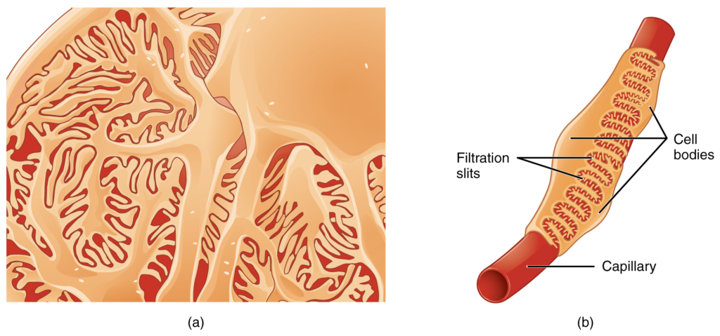
Podocytes interdigitate with structures called pedicels and filter substances in a way similar to fenestrations. In (a), the large cell body can be seen at the top right corner, with branches extending from the cell body. The smallest finger-like extensions are the pedicels. Pedicels on one podocyte always interdigitate with the pedicels of another podocyte. (b) This capillary has three podocytes wrapped around it.

Fenestrations allow many substances to diffuse from the blood based primarily on size.

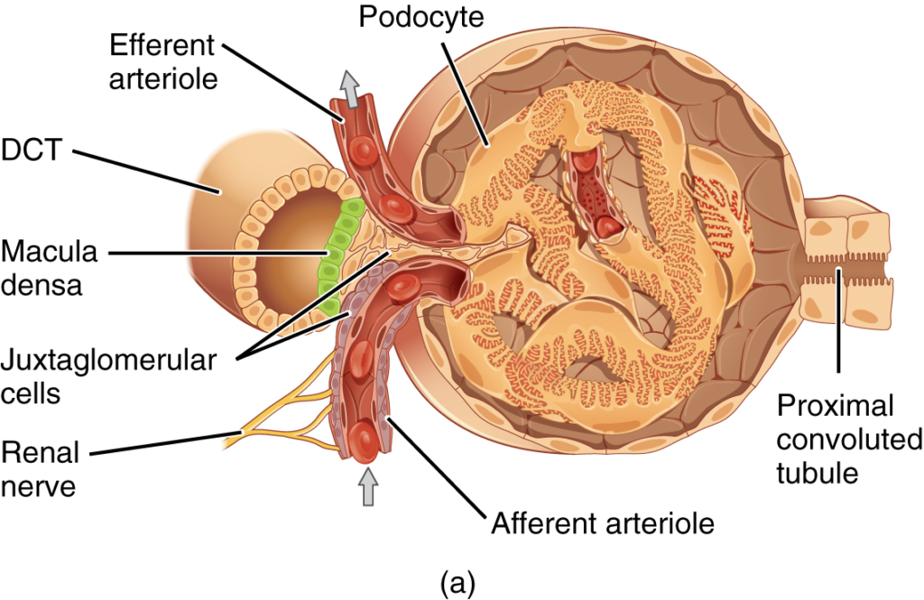
(a) The JGA allows specialized cells to monitor the composition of the fluid in the DCT and adjust the glomerular filtration rate. (b) This micrograph shows the glomerulus and surrounding structures. LM × 1540. (Micrograph provided by the Regents of University of Michigan Medical School © 2012)

The enzyme renin converts the pro-enzyme angiotensin I; the lung-derived enzyme ACE converts angiotensin I into active angiotensin II.

Positive charges inside the channel prevent the leakage of electrolytes across the cell membrane, while allowing water to move due to osmosis.

Nội dung này đang được cập nhật.
Dưới đây là video và các luyện tập (practice) của bài này. Nghe là một kĩ năng khó, đặc biệt là khi chúng ta chưa quen nội dung và chưa có nhạy cảm ngôn ngữ. Nhưng cứ đi thật chậm và đừng bỏ cuộc.
Xem video và cảm nhận nội dung bài. Bạn có thể thả trôi, cảm nhận dòng chảy ngôn ngữ và không nhất thiết phải hiểu toàn bộ bài. Bên dưới là script để bạn khái quát nội dụng và tra từ mới.
Script:
  1. The structure of the kidney is divided into two principle regions—the peripheral rim of cortex and the central medulla.
  2. The two kidneys receive about 25 percent of cardiac output.
  3. Kidney function is derived from the actions of about 1.3 million nephrons per kidney; these are the “functional units.”
  4. The functional unit of the kidney, the nephron, consists of the renal corpuscle, proximal convoluted tubule, loop of Henle, and distal convoluted tubule.
  5. Cortical nephrons have short loops of Henle, whereas juxtamedullary nephrons have long loops of Henle extending into the medulla.
  6. About 15 percent of nephrons are juxtamedullary.
  7. The glomerulus is a capillary bed that filters blood principally based on particle size.
  8. The filtrate is captured by Bowman’s capsule and directed to the proximal convoluted tubule.
  9. A filtration membrane is formed by the fused basement membranes of the podocytes and the capillary endothelial cells that they embrace.
  10. Contractile mesangial cells further perform a role in regulating the rate at which the blood is filtered.
  11. Specialized cells in the juxtaglomerular apparatus produce paracrine signals to regulate blood flow and filtration rates of the glomerulus.
  12. Other juxtaglomerular apparatus cells produce the enzyme renin, which plays a central role in blood pressure regulation.
  13. The filtrate enters the proximal convoluted tubule where absorption and secretion of several substances occur.
  14. The descending and ascending limbs of the loop of Henle consist of thick and thin segments.
  15. Absorption and secretion continue in the distal convoluted tubule but to a lesser extent than in the proximal convoluted tubule.
  16. Each collecting duct collects forming urine from several nephrons and responds to the posterior pituitary hormone antidiuretic hormone by inserting aquaporin water channels into the cell membrane to fine tune water recovery.
Bật video, nghe và điền từ vào chỗ trống.
Dưới đây là phần bàn luận. Bạn có thể tự do đặt câu hỏi, bổ sung kiến thức, và chia sẻ trải nghiệm của mình.
Subscribe
Notify of

0 Comments
Inline Feedbacks
View all comments

Ấn vào ô bên dưới để đánh dấu bạn đã hoàn thành bài học này

Quá dữ! Tiếp tục duy trì phong độ nhé!