Module 14: The Cardiovascular System: The Heart

Lesson 2: Heart Anatomy: Internal Structures

Giải Phẫu Tim: Cấu Trúc Bên Trong

Nội dung bài học:
Mỗi bài học (lesson) bao gồm 4 phần chính: Thuật ngữ, Luyện Đọc, Luyện Nghe, và Bàn Luận.
Sử dụng tính năng:
Bôi hoặc nhấp đôi vào từ, sau đó ấn vào biểu tượng để tra nghĩa từ đó. Khi bạn đưa chuột đến câu (hoặc khi nhấp vào câu trên màn hình điện thoại), gợi ý về cách hiểu câu đó sẽ hiện lên. Cuối cùng, bạn có thể đánh dấu hoàn thành toàn bộ bài học bằng cách ấn vào nút “Hoàn Thành” ở cuối trang.
Đăng ký và đăng nhập
Bạn cần đăng ký và đăng nhập vào tài khoản để lưu quá trình học.
Dưới đây là danh sách những thuật ngữ Y khoa của module The Cardiovascular System: The Heart.
Khái quát được số lượng thuật ngữ sẽ xuất hiện trong bài đọc và nghe sẽ giúp bạn thoải mái tiêu thụ nội dung hơn. Sau khi hoàn thành nội dung đọc và nghe, bạn hãy quay lại đây và luyện tập (practice) để quen dần các thuật ngữ này. Đừng ép bản thân phải nhớ các thuật ngữ này vội vì bạn sẽ gặp và ôn lại danh sách này trong những bài học (lesson) khác của cùng một module.

Medical Terminology: The Cardiovascular System: The Heart

afterload
force the ventricles must develop to effectively pump blood against the resistance in the vessels
anastomosis
(plural = anastomoses) area where vessels unite to allow blood to circulate even if there may be partial blockage in another branch
anterior cardiac veins
vessels that parallel the small cardiac arteries and drain the anterior surface of the right ventricle; bypass the coronary sinus and drain directly into the right atrium
anterior interventricular artery
(also, left anterior descending artery or LAD) major branch of the left coronary artery that follows the anterior interventricular sulcus
anterior interventricular sulcus
sulcus located between the left and right ventricles on the anterior surface of the heart
aortic valve
(also, aortic semilunar valve) valve located at the base of the aorta
artificial pacemaker
medical device that transmits electrical signals to the heart to ensure that it contracts and pumps blood to the body
atrial reflex
(also, called Bainbridge reflex) autonomic reflex that responds to stretch receptors in the atria that send impulses to the cardioaccelerator area to increase HR when venous flow into the atria increases
atrioventricular (AV) node
clump of myocardial cells located in the inferior portion of the right atrium within the atrioventricular septum; receives the impulse from the SA node, pauses, and then transmits it into specialized conducting cells within the interventricular septum
atrioventricular bundle
(also, bundle of His) group of specialized myocardial conductile cells that transmit the impulse from the AV node through the interventricular septum; form the left and right atrioventricular bundle branches
atrioventricular bundle branches
(also, left or right bundle branches) specialized myocardial conductile cells that arise from the bifurcation of the atrioventricular bundle and pass through the interventricular septum; lead to the Purkinje fibers and also to the right papillary muscle via the moderator band
atrioventricular septum
cardiac septum located between the atria and ventricles; atrioventricular valves are located here
atrioventricular valves
one-way valves located between the atria and ventricles; the valve on the right is called the tricuspid valve, and the one on the left is the mitral or bicuspid valve
atrium
(plural = atria) upper or receiving chamber of the heart that pumps blood into the lower chambers just prior to their contraction; the right atrium receives blood from the systemic circuit that flows into the right ventricle; the left atrium receives blood from the pulmonary circuit that flows into the left ventricle
auricle
extension of an atrium visible on the superior surface of the heart
autonomic tone
contractile state during resting cardiac activity produced by mild sympathetic and parasympathetic stimulation
autorhythmicity
ability of cardiac muscle to initiate its own electrical impulse that triggers the mechanical contraction that pumps blood at a fixed pace without nervous or endocrine control
Bachmann’s bundle
(also, interatrial band) group of specialized conducting cells that transmit the impulse directly from the SA node in the right atrium to the left atrium
Bainbridge reflex
(also, called atrial reflex) autonomic reflex that responds to stretch receptors in the atria that send impulses to the cardioaccelerator area to increase HR when venous flow into the atria increases
baroreceptor reflex
autonomic reflex in which the cardiac centers monitor signals from the baroreceptor stretch receptors and regulate heart function based on blood flow
bicuspid valve
(also, mitral valve or left atrioventricular valve) valve located between the left atrium and ventricle; consists of two flaps of tissue
bulbus cordis
portion of the primitive heart tube that will eventually develop into the right ventricle
bundle of His
(also, atrioventricular bundle) group of specialized myocardial conductile cells that transmit the impulse from the AV node through the interventricular septum; form the left and right atrioventricular bundle branches
cardiac cycle
period of time between the onset of atrial contraction (atrial systole) and ventricular relaxation (ventricular diastole)
cardiac notch
depression in the medial surface of the superior lobe of the left lung where the apex of the heart is located
cardiac output (CO)
amount of blood pumped by each ventricle during one minute; equals HR multiplied by SV
cardiac plexus
paired complex network of nerve fibers near the base of the heart that receive sympathetic and parasympathetic stimulations to regulate HR
cardiac reflexes
series of autonomic reflexes that enable the cardiovascular centers to regulate heart function based upon sensory information from a variety of visceral sensors
cardiac reserve
difference between maximum and resting CO
cardiac skeleton
(also, skeleton of the heart) reinforced connective tissue located within the atrioventricular septum; includes four rings that surround the openings between the atria and ventricles, and the openings to the pulmonary trunk and aorta; the point of attachment for the heart valves
cardiogenic area
area near the head of the embryo where the heart begins to develop 18–19 days after fertilization
cardiogenic cords
two strands of tissue that form within the cardiogenic area
cardiomyocyte
muscle cell of the heart
chordae tendineae
string-like extensions of tough connective tissue that extend from the flaps of the atrioventricular valves to the papillary muscles
circumflex artery
branch of the left coronary artery that follows coronary sulcus
coronary arteries
branches of the ascending aorta that supply blood to the heart; the left coronary artery feeds the left side of the heart, the left atrium and ventricle, and the interventricular septum; the right coronary artery feeds the right atrium, portions of both ventricles, and the heart conduction system
coronary sinus
large, thin-walled vein on the posterior surface of the heart that lies within the atrioventricular sulcus and drains the heart myocardium directly into the right atrium
coronary sulcus
sulcus that marks the boundary between the atria and ventricles
coronary veins
vessels that drain the heart and generally parallel the large surface arteries
diastole
period of time when the heart muscle is relaxed and the chambers fill with blood
ejection fraction
portion of the blood that is pumped or ejected from the heart with each contraction; mathematically represented by SV divided by EDV
electrocardiogram (ECG)
surface recording of the electrical activity of the heart that can be used for diagnosis of irregular heart function; also abbreviated as EKG
end diastolic volume (EDV)
(also, preload) the amount of blood in the ventricles at the end of atrial systole just prior to ventricular contraction
end systolic volume (ESV)
amount of blood remaining in each ventricle following systole
endocardial tubes
stage in which lumens form within the expanding cardiogenic cords, forming hollow structures
endocardium
innermost layer of the heart lining the heart chambers and heart valves; composed of endothelium reinforced with a thin layer of connective tissue that binds to the myocardium
endothelium
layer of smooth, simple squamous epithelium that lines the endocardium and blood vessels
epicardial coronary arteries
surface arteries of the heart that generally follow the sulci
epicardium
innermost layer of the serous pericardium and the outermost layer of the heart wall
filling time
duration of ventricular diastole during which filling occurs
foramen ovale
opening in the fetal heart that allows blood to flow directly from the right atrium to the left atrium, bypassing the fetal pulmonary circuit
fossa ovalis
oval-shaped depression in the interatrial septum that marks the former location of the foramen ovale
Frank-Starling mechanism
relationship between ventricular stretch and contraction in which the force of heart contraction is directly proportional to the initial length of the muscle fiber
great cardiac vein
vessel that follows the interventricular sulcus on the anterior surface of the heart and flows along the coronary sulcus into the coronary sinus on the posterior surface; parallels the anterior interventricular artery and drains the areas supplied by this vessel
heart block
interruption in the normal conduction pathway
heart bulge
prominent feature on the anterior surface of the heart, reflecting early cardiac development
heart rate (HR)
number of times the heart contracts (beats) per minute
heart sounds
sounds heard via auscultation with a stethoscope of the closing of the atrioventricular valves (“lub”) and semilunar valves (“dub”)
hypertrophic cardiomyopathy
pathological enlargement of the heart, generally for no known reason
inferior vena cava
large systemic vein that returns blood to the heart from the inferior portion of the body
interatrial band
(also, Bachmann’s bundle) group of specialized conducting cells that transmit the impulse directly from the SA node in the right atrium to the left atrium
interatrial septum
cardiac septum located between the two atria; contains the fossa ovalis after birth
intercalated disc
physical junction between adjacent cardiac muscle cells; consisting of desmosomes, specialized linking proteoglycans, and gap junctions that allow passage of ions between the two cells
internodal pathways
specialized conductile cells within the atria that transmit the impulse from the SA node throughout the myocardial cells of the atrium and to the AV node
interventricular septum
cardiac septum located between the two ventricles
isovolumic contraction
(also, isovolumetric contraction) initial phase of ventricular contraction in which tension and pressure in the ventricle increase, but no blood is pumped or ejected from the heart
isovolumic ventricular relaxation phase
initial phase of the ventricular diastole when pressure in the ventricles drops below pressure in the two major arteries, the pulmonary trunk, and the aorta, and blood attempts to flow back into the ventricles, producing the dicrotic notch of the ECG and closing the two semilunar valves
left atrioventricular valve
(also, mitral valve or bicuspid valve) valve located between the left atrium and ventricle; consists of two flaps of tissue
marginal arteries
branches of the right coronary artery that supply blood to the superficial portions of the right ventricle
mesoderm
one of the three primary germ layers that differentiate early in embryonic development
mesothelium
simple squamous epithelial portion of serous membranes, such as the superficial portion of the epicardium (the visceral pericardium) and the deepest portion of the pericardium (the parietal pericardium)
middle cardiac vein
vessel that parallels and drains the areas supplied by the posterior interventricular artery; drains into the great cardiac vein
mitral valve
(also, left atrioventricular valve or bicuspid valve) valve located between the left atrium and ventricle; consists of two flaps of tissue
moderator band
band of myocardium covered by endocardium that arises from the inferior portion of the interventricular septum in the right ventricle and crosses to the anterior papillary muscle; contains conductile fibers that carry electrical signals followed by contraction of the heart
murmur
unusual heart sound detected by auscultation; typically related to septal or valve defects
myocardial conducting cells
specialized cells that transmit electrical impulses throughout the heart and trigger contraction by the myocardial contractile cells
myocardial contractile cells
bulk of the cardiac muscle cells in the atria and ventricles that conduct impulses and contract to propel blood
myocardium
thickest layer of the heart composed of cardiac muscle cells built upon a framework of primarily collagenous fibers and blood vessels that supply it and the nervous fibers that help to regulate it
negative inotropic factors
factors that negatively impact or lower heart contractility
P wave
component of the electrocardiogram that represents the depolarization of the atria
pacemaker
cluster of specialized myocardial cells known as the SA node that initiates the sinus rhythm
papillary muscle
extension of the myocardium in the ventricles to which the chordae tendineae attach
pectinate muscles
muscular ridges seen on the anterior surface of the right atrium
pericardial cavity
cavity surrounding the heart filled with a lubricating serous fluid that reduces friction as the heart contracts
pericardial sac
(also, pericardium) membrane that separates the heart from other mediastinal structures; consists of two distinct, fused sublayers: the fibrous pericardium and the parietal pericardium
pericardium
(also, pericardial sac) membrane that separates the heart from other mediastinal structures; consists of two distinct, fused sublayers: the fibrous pericardium and the parietal pericardium
positive inotropic factors
factors that positively impact or increase heart contractility
posterior cardiac vein
vessel that parallels and drains the areas supplied by the marginal artery branch of the circumflex artery; drains into the great cardiac vein
posterior interventricular artery
(also, posterior descending artery) branch of the right coronary artery that runs along the posterior portion of the interventricular sulcus toward the apex of the heart and gives rise to branches that supply the interventricular septum and portions of both ventricles
posterior interventricular sulcus
sulcus located between the left and right ventricles on the posterior surface of the heart
preload
(also, end diastolic volume) amount of blood in the ventricles at the end of atrial systole just prior to ventricular contraction
prepotential depolarization
(also, spontaneous depolarization) mechanism that accounts for the autorhythmic property of cardiac muscle; the membrane potential increases as sodium ions diffuse through the always-open sodium ion channels and causes the electrical potential to rise
primitive atrium
portion of the primitive heart tube that eventually becomes the anterior portions of both the right and left atria, and the two auricles
primitive heart tube
singular tubular structure that forms from the fusion of the two endocardial tubes
primitive ventricle
portion of the primitive heart tube that eventually forms the left ventricle
pulmonary arteries
left and right branches of the pulmonary trunk that carry deoxygenated blood from the heart to each of the lungs
pulmonary capillaries
capillaries surrounding the alveoli of the lungs where gas exchange occurs: carbon dioxide exits the blood and oxygen enters
pulmonary circuit
blood flow to and from the lungs
pulmonary trunk
large arterial vessel that carries blood ejected from the right ventricle; divides into the left and right pulmonary arteries
pulmonary valve
(also, pulmonary semilunar valve, the pulmonic valve, or the right semilunar valve) valve at the base of the pulmonary trunk that prevents backflow of blood into the right ventricle; consists of three flaps
pulmonary veins
veins that carry highly oxygenated blood into the left atrium, which pumps the blood into the left ventricle, which in turn pumps oxygenated blood into the aorta and to the many branches of the systemic circuit
Purkinje fibers
specialized myocardial conduction fibers that arise from the bundle branches and spread the impulse to the myocardial contraction fibers of the ventricles
QRS complex
component of the electrocardiogram that represents the depolarization of the ventricles and includes, as a component, the repolarization of the atria
right atrioventricular valve
(also, tricuspid valve) valve located between the right atrium and ventricle; consists of three flaps of tissue
semilunar valves
valves located at the base of the pulmonary trunk and at the base of the aorta
septum
(plural = septa) walls or partitions that divide the heart into chambers
septum primum
flap of tissue in the fetus that covers the foramen ovale within a few seconds after birth
sinoatrial (SA) node
known as the pacemaker, a specialized clump of myocardial conducting cells located in the superior portion of the right atrium that has the highest inherent rate of depolarization that then spreads throughout the heart
sinus rhythm
normal contractile pattern of the heart
sinus venosus
develops into the posterior portion of the right atrium, the SA node, and the coronary sinus
small cardiac vein
parallels the right coronary artery and drains blood from the posterior surfaces of the right atrium and ventricle; drains into the coronary sinus, middle cardiac vein, or right atrium
spontaneous depolarization
(also, prepotential depolarization) the mechanism that accounts for the autorhythmic property of cardiac muscle; the membrane potential increases as sodium ions diffuse through the always-open sodium ion channels and causes the electrical potential to rise
stroke volume (SV)
amount of blood pumped by each ventricle per contraction; also, the difference between EDV and ESV
sulcus
(plural = sulci) fat-filled groove visible on the surface of the heart; coronary vessels are also located in these areas
superior vena cava
large systemic vein that returns blood to the heart from the superior portion of the body
systemic circuit
blood flow to and from virtually all of the tissues of the body
systole
period of time when the heart muscle is contracting
T wave
component of the electrocardiogram that represents the repolarization of the ventricles
target heart rate
range in which both the heart and lungs receive the maximum benefit from an aerobic workout
trabeculae carneae
ridges of muscle covered by endocardium located in the ventricles
tricuspid valve
term used most often in clinical settings for the right atrioventricular valve
truncus arteriosus
portion of the primitive heart that will eventually divide and give rise to the ascending aorta and pulmonary trunk
valve
in the cardiovascular system, a specialized structure located within the heart or vessels that ensures one-way flow of blood
ventricle
one of the primary pumping chambers of the heart located in the lower portion of the heart; the left ventricle is the major pumping chamber on the lower left side of the heart that ejects blood into the systemic circuit via the aorta and receives blood from the left atrium; the right ventricle is the major pumping chamber on the lower right side of the heart that ejects blood into the pulmonary circuit via the pulmonary trunk and receives blood from the right atrium
ventricular ejection phase
second phase of ventricular systole during which blood is pumped from the ventricle
Nội dung này đang được cập nhật.
Dưới đây là các bài văn nằm ở bên trái. Ở bên phải là các bài luyện tập (practice) để đánh giá khả năng đọc hiểu của bạn. Sẽ khó khăn trong thời gian đầu nếu vốn từ vựng của bạn còn hạn chế, đặc biệt là từ vựng Y khoa. Hãy kiên nhẫn và đọc nhiều nhất có kể, lượng kiến thức tích tụ dần sẽ giúp bạn đọc thoải mái hơn.
Recall that the heart’s contraction cycle follows a dual pattern of circulation—the pulmonary and systemic circuits—because of the pairs of chambers that pump blood into the circulation. In order to develop a more precise understanding of cardiac function, it is first necessary to explore the internal anatomical structures in more detail.
The word septum is derived from the Latin for “something that encloses;” in this case, a septum (plural = septa) refers to a wall or partition that divides the heart into chambers. The septa are physical extensions of the myocardium lined with endocardium. Located between the two atria is the interatrial septum. Normally in an adult heart, the interatrial septum bears an oval-shaped depression known as the fossa ovalis, a remnant of an opening in the fetal heart known as the foramen ovale. The foramen ovale allowed blood in the fetal heart to pass directly from the right atrium to the left atrium, allowing some blood to bypass the pulmonary circuit. Within seconds after birth, a flap of tissue known as the septum primum that previously acted as a valve closes the foramen ovale and establishes the typical cardiac circulation pattern.

Between the two ventricles is a second septum known as the interventricular septum. Unlike the interatrial septum, the interventricular septum is normally intact after its formation during fetal development. It is substantially thicker than the interatrial septum, since the ventricles generate far greater pressure when they contract.

The septum between the atria and ventricles is known as the atrioventricular septum. It is marked by the presence of four openings that allow blood to move from the atria into the ventricles and from the ventricles into the pulmonary trunk and aorta. Located in each of these openings between the atria and ventricles is a valve, a specialized structure that ensures one-way flow of blood. The valves between the atria and ventricles are known generically as atrioventricular valves. The valves at the openings that lead to the pulmonary trunk and aorta are known generically as semilunar valves. The interventricular septum is visible in Figure 1. In this figure, the atrioventricular septum has been removed to better show the bicuspid and tricuspid valves; the interatrial septum is not visible, since its location is covered by the aorta and pulmonary trunk. Since these openings and valves structurally weaken the atrioventricular septum, the remaining tissue is heavily reinforced with dense connective tissue called the cardiac skeleton, or skeleton of the heart. It includes four rings that surround the openings between the atria and ventricles, and the openings to the pulmonary trunk and aorta, and serve as the point of attachment for the heart valves. The cardiac skeleton also provides an important boundary in the heart electrical conduction system.
The right atrium serves as the receiving chamber for blood returning to the heart from the systemic circulation. The two major systemic veins, the superior and inferior venae cavae, and the large coronary vein called the coronary sinus that drains the heart myocardium empty into the right atrium. The superior vena cava drains blood from regions superior to the diaphragm: the head, neck, upper limbs, and the thoracic region. It empties into the superior and posterior portions of the right atrium. The inferior vena cava drains blood from areas inferior to the diaphragm: the lower limbs and abdominopelvic region of the body. It, too, empties into the posterior portion of the atria, but inferior to the opening of the superior vena cava. Immediately superior and slightly medial to the opening of the inferior vena cava on the posterior surface of the atrium is the opening of the coronary sinus. This thin-walled vessel drains most of the coronary veins that return systemic blood from the heart. The majority of the internal heart structures discussed in this and subsequent sections are illustrated in Figure 1.

While the bulk of the internal surface of the right atrium is smooth, the depression of the fossa ovalis is medial, and the anterior surface demonstrates prominent ridges of muscle called the pectinate muscles. The right auricle also has pectinate muscles. The left atrium does not have pectinate muscles except in the auricle.

The atria receive venous blood on a nearly continuous basis, preventing venous flow from stopping while the ventricles are contracting. While most ventricular filling occurs while the atria are relaxed, they do demonstrate a contractile phase and actively pump blood into the ventricles just prior to ventricular contraction. The opening between the atrium and ventricle is guarded by the tricuspid valve.
The right ventricle receives blood from the right atrium through the tricuspid valve. Each flap of the valve is attached to strong strands of connective tissue, the chordae tendineae, literally “tendinous cords,” or sometimes more poetically referred to as “heart strings.” There are several chordae tendineae associated with each of the flaps. They are composed of approximately 80 percent collagenous fibers with the remainder consisting of elastic fibers and endothelium. They connect each of the flaps to a papillary muscle that extends from the inferior ventricular surface. There are three papillary muscles in the right ventricle, called the anterior, posterior, and septal muscles, which correspond to the three sections of the valves.

When the myocardium of the ventricle contracts, pressure within the ventricular chamber rises. Blood, like any fluid, flows from higher pressure to lower pressure areas, in this case, toward the pulmonary trunk and the atrium. To prevent any potential backflow, the papillary muscles also contract, generating tension on the chordae tendineae. This prevents the flaps of the valves from being forced into the atria and regurgitation of the blood back into the atria during ventricular contraction. Figure 2 shows papillary muscles and chordae tendineae attached to the tricuspid valve.

The walls of the ventricle are lined with trabeculae carneae, ridges of cardiac muscle covered by endocardium. In addition to these muscular ridges, a band of cardiac muscle, also covered by endocardium, known as the moderator band (see Figure 1) reinforces the thin walls of the right ventricle and plays a crucial role in cardiac conduction. It arises from the inferior portion of the interventricular septum and crosses the interior space of the right ventricle to connect with the inferior papillary muscle.

When the right ventricle contracts, it ejects blood into the pulmonary trunk, which branches into the left and right pulmonary arteries that carry it to each lung. The superior surface of the right ventricle begins to taper as it approaches the pulmonary trunk. At the base of the pulmonary trunk is the pulmonary semilunar valve that prevents backflow from the pulmonary trunk.
After exchange of gases in the pulmonary capillaries, blood returns to the left atrium high in oxygen via one of the four pulmonary veins. While the left atrium does not contain pectinate muscles, it does have an auricle that includes these pectinate ridges. Blood flows nearly continuously from the pulmonary veins back into the atrium, which acts as the receiving chamber, and from here through an opening into the left ventricle. Most blood flows passively into the heart while both the atria and ventricles are relaxed, but toward the end of the ventricular relaxation period, the left atrium will contract, pumping blood into the ventricle. This atrial contraction accounts for approximately 20 percent of ventricular filling. The opening between the left atrium and ventricle is guarded by the mitral valve.
Recall that, although both sides of the heart will pump the same amount of blood, the muscular layer is much thicker in the left ventricle compared to the right (see Figure 3). Like the right ventricle, the left also has trabeculae carneae, but there is no moderator band. The mitral valve is connected to papillary muscles via chordae tendineae. There are two papillary muscles on the left—the anterior and posterior—as opposed to three on the right.

The left ventricle is the major pumping chamber for the systemic circuit; it ejects blood into the aorta through the aortic semilunar valve.
A transverse section through the heart slightly above the level of the atrioventricular septum reveals all four heart valves along the same plane (Figure 4). The valves ensure unidirectional blood flow through the heart. Between the right atrium and the right ventricle is the right atrioventricular valve, or tricuspid valve. It typically consists of three flaps, or leaflets, made of endocardium reinforced with additional connective tissue. The flaps are connected by chordae tendineae to the papillary muscles, which control the opening and closing of the valves.

Emerging from the right ventricle at the base of the pulmonary trunk is the pulmonary semilunar valve, or the pulmonary valve; it is also known as the pulmonic valve or the right semilunar valve. The pulmonary valve is comprised of three small flaps of endothelium reinforced with connective tissue. When the ventricle relaxes, the pressure differential causes blood to flow back into the ventricle from the pulmonary trunk. This flow of blood fills the pocket-like flaps of the pulmonary valve, causing the valve to close and producing an audible sound. Unlike the atrioventricular valves, there are no papillary muscles or chordae tendineae associated with the pulmonary valve.

Located at the opening between the left atrium and left ventricle is the mitral valve, also called the bicuspid valve or the left atrioventricular valve. Structurally, this valve consists of two cusps, compared to the three cusps of the tricuspid valve. In a clinical setting, the valve is referred to as the mitral valve, rather than the bicuspid valve. The two cusps of the mitral valve are attached by chordae tendineae to two papillary muscles that project from the wall of the ventricle.

At the base of the aorta is the aortic semilunar valve, or the aortic valve, which prevents backflow from the aorta. It normally is composed of three flaps. When the ventricle relaxes and blood attempts to flow back into the ventricle from the aorta, blood will fill the cusps of the valve, causing it to close and producing an audible sound.

In Figure 5a, the two atrioventricular valves are open and the two semilunar valves are closed. This occurs when both atria and ventricles are relaxed and when the atria contract to pump blood into the ventricles. Figure 5b shows a frontal view. Although only the left side of the heart is illustrated, the process is virtually identical on the right.

Figure 6a shows the atrioventricular valves closed while the two semilunar valves are open. This occurs when the ventricles contract to eject blood into the pulmonary trunk and aorta. Closure of the two atrioventricular valves prevents blood from being forced back into the atria. This stage can be seen from a frontal view in Figure 6b.

OpenStax. (2022). Anatomy and Physiology 2e. Rice University. Retrieved June 15, 2023. ISBN-13: 978-1-711494-06-7 (Hardcover) ISBN-13: 978-1-711494-05-0 (Paperback) ISBN-13: 978-1-951693-42-8 (Digital). License: Attribution 4.0 International (CC BY 4.0). Access for free at openstax.org.

This anterior view of the heart shows the four chambers, the major vessels and their early branches, as well as the valves. The presence of the pulmonary trunk and aorta covers the interatrial septum, and the atrioventricular septum is cut away to show the atrioventricular valves.

In this frontal section, you can see papillary muscles attached to the tricuspid valve on the right as well as the mitral valve on the left via chordae tendineae. (credit: modification of work by “PV KS”/flickr.com)

The myocardium in the left ventricle is significantly thicker than that of the right ventricle. Both ventricles pump the same amount of blood, but the left ventricle must generate a much greater pressure to overcome greater resistance in the systemic circuit. The ventricles are shown in both relaxed and contracting states. Note the differences in the relative size of the lumens, the region inside each ventricle where the blood is contained.

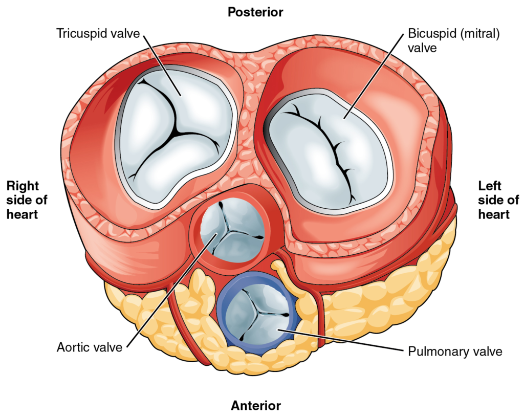
With the atria and major vessels removed, all four valves are clearly visible, although it is difficult to distinguish the three separate cusps of the tricuspid valve.

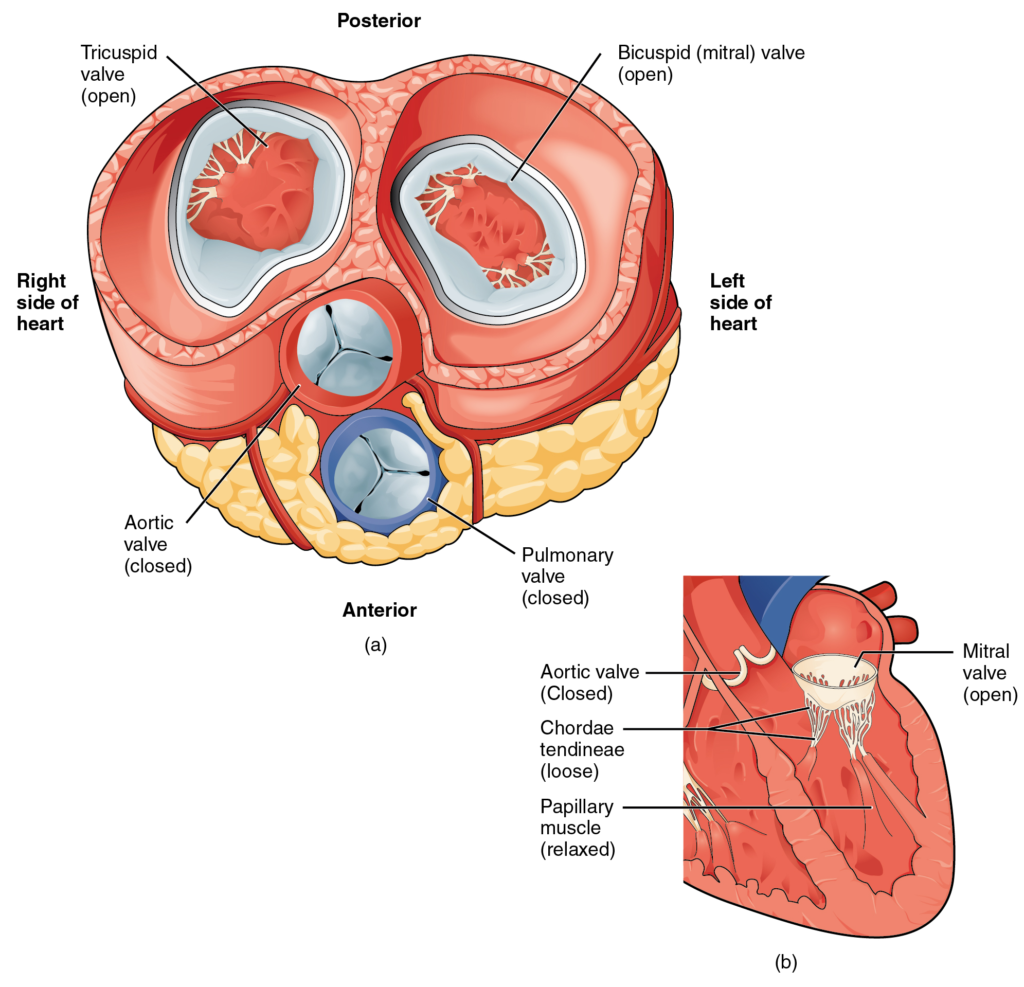
(a) A transverse section through the heart illustrates the four heart valves. The two atrioventricular valves are open; the two semilunar valves are closed. The atria and vessels have been removed. (b) A frontal section through the heart illustrates blood flow through the mitral valve. When the mitral valve is open, it allows blood to move from the left atrium to the left ventricle. The aortic semilunar valve is closed to prevent backflow of blood from the aorta to the left ventricle.

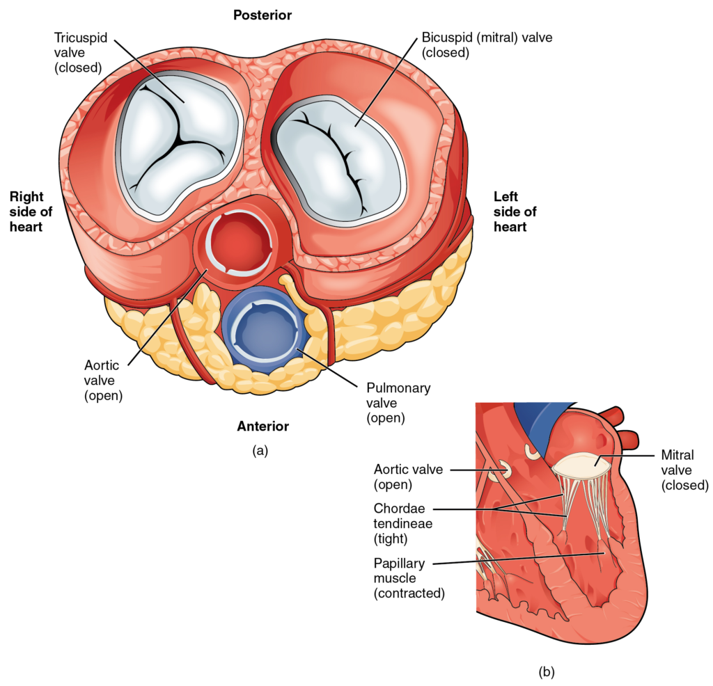
(a) A transverse section through the heart illustrates the four heart valves during ventricular contraction. The two atrioventricular valves are closed, but the two semilunar valves are open. The atria and vessels have been removed. (b) A frontal view shows the closed mitral (bicuspid) valve that prevents backflow of blood into the left atrium. The aortic semilunar valve is open to allow blood to be ejected into the aorta.

Nội dung này đang được cập nhật.
Dưới đây là video và các luyện tập (practice) của bài này. Nghe là một kĩ năng khó, đặc biệt là khi chúng ta chưa quen nội dung và chưa có nhạy cảm ngôn ngữ. Nhưng cứ đi thật chậm và đừng bỏ cuộc.
Xem video và cảm nhận nội dung bài. Bạn có thể thả trôi, cảm nhận dòng chảy ngôn ngữ và không nhất thiết phải hiểu toàn bộ bài. Bên dưới là script để bạn khái quát nội dụng và tra từ mới.
Script:
  1. The heart resides within the pericardial sac and is located in the mediastinal space within the thoracic cavity.
  2. The pericardial sac consists of two fused layers: an outer fibrous capsule and an inner parietal pericardium lined with a serous membrane.
  3. Between the pericardial sac and the heart is the pericardial cavity, which is filled with lubricating serous fluid.
  4. The walls of the heart are composed of an outer epicardium, a thick myocardium, and an inner lining layer of endocardium.
  5. The human heart consists of a pair of atria, which receive blood and pump it into a pair of ventricles, which pump blood into the vessels.
  6. The right atrium receives systemic blood relatively low in oxygen and pumps it into the right ventricle, which pumps it into the pulmonary circuit.
  7. Exchange of oxygen and carbon dioxide occurs in the lungs.
  8. Then, blood high in oxygen returns to the left atrium, which pumps blood into the left ventricle.
  9. From there, blood is pumped into the aorta and the remainder of the systemic circuit.
  10. The septa are the partitions that separate the chambers of the heart.
  11. They include the interatrial septum, the interventricular septum, and the atrioventricular septum.
  12. Two of these openings are guarded by the atrioventricular valves, the right tricuspid valve and the left mitral valve, which prevent the backflow of blood.
  13. Each is attached to chordae tendineae that extend to the papillary muscles, which are extensions of the myocardium, to prevent the valves from being blown back into the atria.
  14. The pulmonary valve is located at the base of the pulmonary trunk, and the left semilunar valve is located at the base of the aorta.
  15. The right and left coronary arteries are the first to branch off the aorta and arise from two of the three sinuses located near the base of the aorta and are generally located in the sulci.
  16. Cardiac veins parallel the small cardiac arteries and generally drain into the coronary sinus.
Bật video, nghe và điền từ vào chỗ trống.
Dưới đây là phần bàn luận. Bạn có thể tự do đặt câu hỏi, bổ sung kiến thức, và chia sẻ trải nghiệm của mình.
Subscribe
Notify of

0 Comments
Inline Feedbacks
View all comments

Ấn vào ô bên dưới để đánh dấu bạn đã hoàn thành bài học này

Quá dữ! Tiếp tục duy trì phong độ nhé!