Module 27: The Reproductive System

Lesson 3: Anatomy of the Female Reproductive System

Giải Phẫu Hệ Sinh Dục Nữ

Nội dung bài học:
Mỗi bài học (lesson) bao gồm 4 phần chính: Thuật ngữ, Luyện Đọc, Luyện Nghe, và Bàn Luận.
Sử dụng tính năng:
Bôi hoặc nhấp đôi vào từ, sau đó ấn vào biểu tượng để tra nghĩa từ đó. Khi bạn đưa chuột đến câu (hoặc khi nhấp vào câu trên màn hình điện thoại), gợi ý về cách hiểu câu đó sẽ hiện lên. Cuối cùng, bạn có thể đánh dấu hoàn thành toàn bộ bài học bằng cách ấn vào nút “Hoàn Thành” ở cuối trang.
Đăng ký và đăng nhập
Bạn cần đăng ký và đăng nhập vào tài khoản để lưu quá trình học.
Dưới đây là danh sách những thuật ngữ Y khoa của module The Reproductive System.
Khái quát được số lượng thuật ngữ sẽ xuất hiện trong bài đọc và nghe sẽ giúp bạn thoải mái tiêu thụ nội dung hơn. Sau khi hoàn thành nội dung đọc và nghe, bạn hãy quay lại đây và luyện tập (practice) để quen dần các thuật ngữ này. Đừng ép bản thân phải nhớ các thuật ngữ này vội vì bạn sẽ gặp và ôn lại danh sách này trong những bài học (lesson) khác của cùng một module.

Medical Terminology: The Reproductive System

alveoli
(of the breast) milk-secreting cells in the mammary gland
ampulla
(of the uterine tube) middle portion of the uterine tube in which fertilization often occurs
antrum
fluid-filled chamber that characterizes a mature tertiary (antral) follicle
areola
highly pigmented, circular area surrounding the raised nipple and containing areolar glands that secrete fluid important for lubrication during suckling
Bartholin’s glands
(also, greater vestibular glands) glands that produce a thick mucus that maintains moisture in the vulva area; also referred to as the greater vestibular glands
blood–testis barrier
tight junctions between Sertoli cells that prevent bloodborne pathogens from gaining access to later stages of spermatogenesis and prevent the potential for an autoimmune reaction to haploid sperm
body of uterus
middle section of the uterus
broad ligament
wide ligament that supports the uterus by attaching laterally to both sides of the uterus and pelvic wall
bulbourethral glands
(also, Cowper’s glands) glands that secrete a lubricating mucus that cleans and lubricates the urethra prior to and during ejaculation
cervix
elongate inferior end of the uterus where it connects to the vagina
clitoris
(also, glans clitoris) nerve-rich area of the vulva that contributes to sexual sensation during intercourse
corpus albicans
nonfunctional structure remaining in the ovarian stroma following structural and functional regression of the corpus luteum
corpus cavernosum
(plural = corpora cavernosa) either of two columns of erectile tissue in the penis that fill with blood during an erection
corpus luteum
transformed follicle after ovulation that secretes progesterone
corpus spongiosum
column of erectile tissue in the penis that fills with blood during an erection and surrounds the penile urethra on the ventral portion of the penis
ductus deferens
(also, vas deferens) duct that transports sperm from the epididymis through the spermatic cord and into the ejaculatory duct; also referred as the vas deferens
ejaculatory duct
duct that connects the ampulla of the ductus deferens with the duct of the seminal vesicle at the prostatic urethra
endometrium
inner lining of the uterus, part of which builds up during the secretory phase of the menstrual cycle and then sheds with menses
epididymis
(plural = epididymides) coiled tubular structure in which sperm start to mature and are stored until ejaculation
fimbriae
fingerlike projections on the distal uterine tubes
follicle
ovarian structure of one oocyte and surrounding granulosa (and later theca) cells
folliculogenesis
development of ovarian follicles from primordial to tertiary under the stimulation of gonadotropins
fundus
(of the uterus) domed portion of the uterus that is superior to the uterine tubes
gamete
haploid reproductive cell that contributes genetic material to form an offspring
glans penis
bulbous end of the penis that contains a large number of nerve endings
gonadotropin-releasing hormone (GnRH)
hormone released by the hypothalamus that regulates the production of follicle-stimulating hormone and luteinizing hormone from the pituitary gland
gonads
reproductive organs (testes and ovaries) that produce gametes and reproductive hormones
granulosa cells
supportive cells in the ovarian follicle that produce estrogen
hymen
membrane that covers part of the opening of the vagina
infundibulum
(of the uterine tube) wide, distal portion of the uterine tube terminating in fimbriae
inguinal canal
opening in abdominal wall that connects the testes to the abdominal cavity
isthmus
narrow, medial portion of the uterine tube that joins the uterus
labia majora
hair-covered folds of skin located behind the mons pubis
labia minora
thin, pigmented, hairless flaps of skin located medial and deep to the labia majora
lactiferous ducts
ducts that connect the mammary glands to the nipple and allow for the transport of milk
lactiferous sinus
area of milk collection between alveoli and lactiferous duct
Leydig cells
cells between the seminiferous tubules of the testes that produce testosterone; a type of interstitial cell
mammary glands
glands inside the breast that secrete milk
menarche
first menstruation in a pubertal female
menses
shedding of the inner portion of the endometrium out though the vagina; also referred to as menstruation
menses phase
phase of the menstrual cycle in which the endometrial lining is shed
menstrual cycle
approximately 28-day cycle of changes in the uterus consisting of a menses phase, a proliferative phase, and a secretory phase
mons pubis
mound of fatty tissue located at the front of the vulva
Müllerian duct
duct system present in the embryo that will eventually form the internal female reproductive structures
myometrium
smooth muscle layer of uterus that allows for uterine contractions during labor and expulsion of menstrual blood
oocyte
cell that results from the division of the oogonium and undergoes meiosis I at the LH surge and meiosis II at fertilization to become a haploid ovum
oogenesis
process by which oogonia divide by mitosis to primary oocytes, which undergo meiosis to produce the secondary oocyte and, upon fertilization, the ovum
oogonia
ovarian stem cells that undergo mitosis during female fetal development to form primary oocytes
ovarian cycle
approximately 28-day cycle of changes in the ovary consisting of a follicular phase and a luteal phase
ovaries
female gonads that produce oocytes and sex steroid hormones (notably estrogen and progesterone)
ovulation
release of a secondary oocyte and associated granulosa cells from an ovary
ovum
haploid female gamete resulting from completion of meiosis II at fertilization
penis
male organ of copulation
perimetrium
outer epithelial layer of uterine wall
polar body
smaller cell produced during the process of meiosis in oogenesis
prepuce
(also, foreskin) flap of skin that forms a collar around, and thus protects and lubricates, the glans penis; also referred as the foreskin
primary follicles
ovarian follicles with a primary oocyte and one layer of cuboidal granulosa cells
primordial follicles
least developed ovarian follicles that consist of a single oocyte and a single layer of flat (squamous) granulosa cells
proliferative phase
phase of the menstrual cycle in which the endometrium proliferates
prostate gland
doughnut-shaped gland at the base of the bladder surrounding the urethra and contributing fluid to semen during ejaculation
puberty
life stage during which a male or female adolescent becomes anatomically and physiologically capable of reproduction
rugae
(of the vagina) folds of skin in the vagina that allow it to stretch during intercourse and childbirth
scrotum
external pouch of skin and muscle that houses the testes
secondary follicles
ovarian follicles with a primary oocyte and multiple layers of granulosa cells
secondary sex characteristics
physical characteristics that are influenced by sex steroid hormones and have supporting roles in reproductive function
secretory phase
phase of the menstrual cycle in which the endometrium secretes a nutrient-rich fluid in preparation for implantation of an embryo
semen
ejaculatory fluid composed of sperm and secretions from the seminal vesicles, prostate, and bulbourethral glands
seminal vesicle
gland that produces seminal fluid, which contributes to semen
seminiferous tubules
tube structures within the testes where spermatogenesis occurs
Sertoli cells
cells that support germ cells through the process of spermatogenesis; a type of sustentacular cell
sperm
(also, spermatozoon) male gamete
spermatic cord
bundle of nerves and blood vessels that supplies the testes; contains ductus deferens
spermatid
immature sperm cells produced by meiosis II of secondary spermatocytes
spermatocyte
cell that results from the division of spermatogonium and undergoes meiosis I and meiosis II to form spermatids
spermatogenesis
formation of new sperm, occurs in the seminiferous tubules of the testes
spermatogonia
(singular = spermatogonium) diploid precursor cells that become sperm
spermiogenesis
transformation of spermatids to spermatozoa during spermatogenesis
suspensory ligaments
bands of connective tissue that suspend the breast onto the chest wall by attachment to the overlying dermis
tertiary follicles
(also, antral follicles) ovarian follicles with a primary or secondary oocyte, multiple layers of granulosa cells, and a fully formed antrum
testes
(singular = testis) male gonads
theca cells
estrogen-producing cells in a maturing ovarian follicle
uterine tubes
(also, fallopian tubes or oviducts) ducts that facilitate transport of an ovulated oocyte to the uterus
uterus
muscular hollow organ in which a fertilized egg develops into a fetus
vagina
tunnel-like organ that provides access to the uterus for the insertion of semen and from the uterus for the birth of a baby
vulva
external female genitalia
Wolffian duct
duct system present in the embryo that will eventually form the internal male reproductive structures
Nội dung này đang được cập nhật.
Dưới đây là các bài văn nằm ở bên trái. Ở bên phải là các bài luyện tập (practice) để đánh giá khả năng đọc hiểu của bạn. Sẽ khó khăn trong thời gian đầu nếu vốn từ vựng của bạn còn hạn chế, đặc biệt là từ vựng Y khoa. Hãy kiên nhẫn và đọc nhiều nhất có kể, lượng kiến thức tích tụ dần sẽ giúp bạn đọc thoải mái hơn.
The female, or ovarian, reproductive system functions to produce gametes and reproductive hormones, just like the male reproductive system; however, it also has the additional task of supporting the developing fetus and delivering it to the outside world. Unlike its male counterpart, the female reproductive system is located primarily inside the pelvic cavity (Figure 1). Recall that the ovaries are the female gonads. The gamete they produce is called an oocyte. We will discuss the production of oocytes in another detailed article. First, let us look at some of the structures of the female reproductive system.
The external female reproductive structures are referred to collectively as the vulva (Figure 2). The mons pubis is a pad of fat that is located at the anterior, over the pubic bone. After puberty, it becomes covered in pubic hair. The labia majora (labia = “lips”; majora = “larger”) are folds of hair-covered skin that begin just posterior to the mons pubis. The thinner and more pigmented labia minora (labia = “lips”; minora = “smaller”) extend medial to the labia majora. Although they naturally vary in shape and size from person to person, the labia minora serve to protect the urethra and the entrance to the reproductive tract.

The superior, anterior portions of the labia minora come together to encircle the clitoris (or glans clitoris), an organ that originates from the same cells as the glans penis and has abundant nerves that make it important in sexual sensation and orgasm. The hymen is a thin membrane that sometimes partially covers the entrance to the vagina. An intact hymen cannot be used as an indication of “virginity”; even at birth, this is only a partial membrane, as menstrual fluid and other secretions must be able to exit the body, regardless of penile–vaginal intercourse. The vaginal opening is located between the opening of the urethra and the anus. It is flanked by outlets to the Bartholin’s glands (or greater vestibular glands).
The vagina, shown at the bottom of Figure 1 and Figure 2, is a muscular canal (approximately 10 cm long) that serves as the entrance to the reproductive tract. It also serves as the exit from the uterus during menses and childbirth. The outer walls of the anterior and posterior vagina are formed into longitudinal columns, or ridges, and the superior portion of the vagina—called the fornix—meets the protruding uterine cervix. The walls of the vagina are lined with an outer, fibrous adventitia; a middle layer of smooth muscle; and an inner mucous membrane with transverse folds called rugae. Together, the middle and inner layers allow the expansion of the vagina to accommodate intercourse and childbirth. The thin, perforated hymen can partially surround the opening to the vaginal orifice. The hymen can be ruptured with strenuous physical exercise, penile–vaginal intercourse, and childbirth. The Bartholin’s glands and the lesser vestibular glands (located near the clitoris) secrete mucus, which keeps the vestibular area moist.

The vagina is home to a normal population of microorganisms that help to protect against infection by pathogenic bacteria, yeast, or other organisms that can enter the vagina. In a healthy person, the most predominant type of vaginal bacteria is from the genus Lactobacillus. This family of beneficial bacterial flora secretes lactic acid, and thus protects the vagina by maintaining an acidic pH (below 4.5). Potential pathogens are less likely to survive in these acidic conditions. Lactic acid, in combination with other vaginal secretions, makes the vagina a self-cleansing organ. However, douching—or washing out the vagina with fluid—can disrupt the normal balance of healthy microorganisms, and actually increase risk for infections and irritation. Indeed, the American College of Obstetricians and Gynecologists recommend that people do not douche, and that they allow the vagina to maintain its normal healthy population of protective microbial flora.
The uterus is the muscular organ that nourishes and supports the growing embryo (Figure 3). Its average size is approximately 5 cm wide by 7 cm long (approximately 2 in by 3 in) when a female is not pregnant. It has three sections. The portion of the uterus superior to the opening of the uterine tubes is called the fundus. The middle section of the uterus is called the body of uterus (or corpus). The cervix is the narrow inferior portion of the uterus that projects into the vagina. The cervix produces mucus secretions that become thin and stringy under the influence of high systemic plasma estrogen concentrations, and these secretions can facilitate sperm movement through the reproductive tract.

Several ligaments maintain the position of the uterus within the abdominopelvic cavity. The broad ligament is a fold of peritoneum that serves as a primary support for the uterus, extending laterally from both sides of the uterus and attaching it to the pelvic wall. The round ligament attaches to the uterus near the uterine tubes, and extends to the labia majora. Finally, the uterosacral ligament stabilizes the uterus posteriorly by its connection from the cervix to the pelvic wall.

The wall of the uterus is made up of three layers. The most superficial layer is the serous membrane, or perimetrium, which consists of epithelial tissue that covers the exterior portion of the uterus. The middle layer, or myometrium, is a thick layer of smooth muscle responsible for uterine contractions. Most of the uterus is myometrial tissue, and the muscle fibers run horizontally, vertically, and diagonally, allowing the powerful contractions that occur during labor and the less powerful contractions (or cramps) that help to expel menstrual blood during a woman’s period. Anteriorly directed myometrial contractions also occur near the time of ovulation, and are thought to possibly facilitate the transport of sperm through the female reproductive tract.

The innermost layer of the uterus is called the endometrium. The endometrium contains a connective tissue lining, the lamina propria, which is covered by epithelial tissue that lines the lumen. Structurally, the endometrium consists of two layers: the stratum basalis and the stratum functionalis (the basal and functional layers). The stratum basalis layer is part of the lamina propria and is adjacent to the myometrium; this layer does not shed during menses. In contrast, the thicker stratum functionalis layer contains the glandular portion of the lamina propria and the endothelial tissue that lines the uterine lumen. It is the stratum functionalis that grows and thickens in response to increased levels of estrogen and progesterone. In the luteal phase of the menstrual cycle, special branches off of the uterine artery called spiral arteries supply the thickened stratum functionalis. This inner functional layer provides the proper site of implantation for the fertilized egg, and—should fertilization not occur—it is only the stratum functionalis layer of the endometrium that sheds during menstruation.

Recall that during the follicular phase of the ovarian cycle, the tertiary follicles are growing and secreting estrogen. At the same time, the stratum functionalis of the endometrium is thickening to prepare for a potential implantation. The post-ovulatory increase in progesterone, which characterizes the luteal phase, is key for maintaining a thick stratum functionalis. As long as a functional corpus luteum is present in the ovary, the endometrial lining is prepared for implantation. Indeed, if an embryo implants, signals are sent to the corpus luteum to continue secreting progesterone to maintain the endometrium, and thus maintain the pregnancy. If an embryo does not implant, no signal is sent to the corpus luteum and it degrades, ceasing progesterone production and ending the luteal phase. Without progesterone, the endometrium thins and, under the influence of prostaglandins, the spiral arteries of the endometrium constrict and rupture, preventing oxygenated blood from reaching the endometrial tissue. As a result, endometrial tissue dies and blood, pieces of the endometrial tissue, and white blood cells are shed through the vagina during menstruation, or the menses. The first menses after puberty, called menarche, can occur either before or after the first ovulation.
The uterine tubes (also called fallopian tubes or oviducts) serve as the conduit of the oocyte from the ovary to the uterus (see Figure 3). Each of the two uterine tubes is close to, but not directly connected to, the ovary and divided into sections. The isthmus is the narrow medial end of each uterine tube that is connected to the uterus. The wide distal infundibulum flares out with slender, finger-like projections called fimbriae. The middle region of the tube, called the ampulla, is where fertilization often occurs. The uterine tubes also have three layers: an outer serosa, a middle smooth muscle layer, and an inner mucosal layer. In addition to its mucus-secreting cells, the inner mucosa contains ciliated cells that beat in the direction of the uterus, producing a current that will be critical to move the oocyte.

Following ovulation, the secondary oocyte surrounded by a few granulosa cells is released into the peritoneal cavity. The nearby uterine tube, either left or right, receives the oocyte. Unlike sperm, oocytes lack flagella, and therefore cannot move on their own. So how do they travel into the uterine tube and toward the uterus? High concentrations of estrogen that occur around the time of ovulation induce contractions of the smooth muscle along the length of the uterine tube. These contractions occur every 4 to 8 seconds, and the result is a coordinated movement that sweeps the surface of the ovary and the pelvic cavity. Current flowing toward the uterus is generated by coordinated beating of the cilia that line the outside and lumen of the length of the uterine tube. These cilia beat more strongly in response to the high estrogen concentrations that occur around the time of ovulation. As a result of these mechanisms, the oocyte–granulosa cell complex is pulled into the interior of the tube. Once inside, the muscular contractions and beating cilia move the oocyte slowly toward the uterus. When fertilization does occur, sperm typically meet the egg while it is still moving through the ampulla.

If the oocyte is successfully fertilized, the resulting zygote will begin to divide into two cells, then four, and so on, as it makes its way through the uterine tube and into the uterus. There, it will implant and continue to grow. If the egg is not fertilized, it will simply degrade—either in the uterine tube or in the uterus, where it may be shed with the next menstrual period.

The open-ended structure of the uterine tubes can have significant health consequences if bacteria or other contagions enter through the vagina and move through the uterus, into the tubes, and then into the pelvic cavity. If this is left unchecked, a bacterial infection (sepsis) could quickly become life-threatening. The spread of an infection in this manner is of special concern when unskilled practitioners perform abortions in non-sterile conditions. Sepsis is also associated with sexually transmitted bacterial infections, especially gonorrhea and chlamydia. These increase a person’s risk for pelvic inflammatory disease (PID), infection of the uterine tubes or other reproductive organs. Even when resolved, PID can leave scar tissue in the tubes, leading to infertility.
The ovaries are the female gonads (see Figure 1). Paired ovals, they are each about 2 to 3 cm in length, about the size of an almond. The ovaries are located within the pelvic cavity, and are supported by the mesovarium, an extension of the peritoneum that connects the ovaries to the broad ligament. Extending from the mesovarium itself is the suspensory ligament that contains the ovarian blood and lymph vessels. Finally, the ovary itself is attached to the uterus via the ovarian ligament.

The ovary comprises an outer covering of cuboidal epithelium called the ovarian surface epithelium that is superficial to a dense connective tissue covering called the tunica albuginea. Beneath the tunica albuginea is the cortex, or outer portion, of the organ. The cortex is composed of a tissue framework called the ovarian stroma that forms the bulk of the adult ovary. Oocytes develop within the outer layer of this stroma, each surrounded by supporting cells. This grouping of an oocyte and its supporting cells is called a follicle. The growth and development of ovarian follicles will be described shortly. Beneath the cortex lies the inner ovarian medulla, the site of blood vessels, lymph vessels, and the nerves of the ovary. You will learn more about the overall anatomy of the female reproductive system at the end of this section.
Whereas the breasts are located far from the other female reproductive organs, they are considered accessory organs of the female reproductive system. The function of the breasts is to supply milk to an infant in a process called lactation. The external features of the breast include a nipple surrounded by a pigmented areola (Figure 4), whose coloration may deepen during pregnancy. The areola is typically circular and can vary in size from 25 to 100 mm in diameter. The areolar region is characterized by small, raised areolar glands that secrete lubricating fluid during lactation to protect the nipple from chafing. When a baby nurses, or draws milk from the breast, the entire areolar region is taken into the mouth.

Breast milk is produced by the mammary glands, which are modified sweat glands. The milk itself exits the breast through the nipple via 15 to 20 lactiferous ducts that open on the surface of the nipple. These lactiferous ducts each extend to a lactiferous sinus that connects to a glandular lobe within the breast itself that contains groups of milk-secreting cells in clusters called alveoli (see Figure 4). The clusters can change in size depending on the amount of milk in the alveolar lumen. Once milk is made in the alveoli, stimulated myoepithelial cells that surround the alveoli contract to push the milk to the lactiferous sinuses. From here, the baby can draw milk through the lactiferous ducts by suckling. The lobes themselves are surrounded by fat tissue, which determines the size of the breast; breast size differs between individuals and does not affect the amount of milk produced. Supporting the breasts are multiple bands of connective tissue called suspensory ligaments that connect the breast tissue to the dermis of the overlying skin.

During the normal hormonal fluctuations in the menstrual cycle, breast tissue responds to changing levels of estrogen and progesterone, which can lead to swelling and breast tenderness in some individuals, especially during the secretory phase. If pregnancy occurs, the increase in hormones leads to further development of the mammary tissue and enlargement of the breasts.

OpenStax. (2022). Anatomy and Physiology 2e. Rice University. Retrieved June 15, 2023. ISBN-13: 978-1-711494-06-7 (Hardcover) ISBN-13: 978-1-711494-05-0 (Paperback) ISBN-13: 978-1-951693-42-8 (Digital). License: Attribution 4.0 International (CC BY 4.0). Access for free at openstax.org.

The major organs of the female reproductive system are located inside the pelvic cavity.

The external female genitalia are referred to collectively as the vulva.

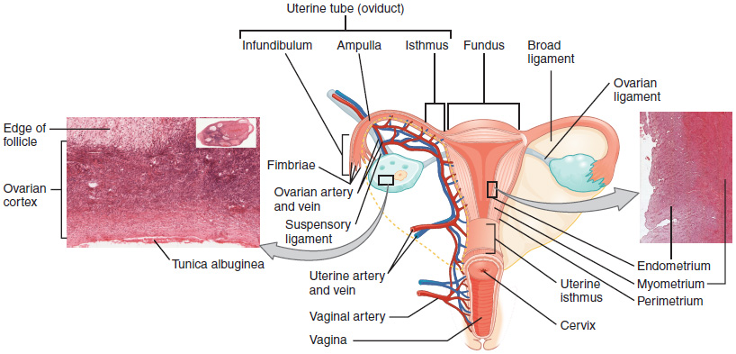
This anterior view shows the relationship of the ovaries, uterine tubes (oviducts), and uterus. Sperm enter through the vagina, and fertilization of an ovulated oocyte usually occurs in the distal uterine tube. From left to right, LM × 400, LM × 20. (Micrographs provided by the Regents of University of Michigan Medical School © 2012)

During lactation, milk moves from the alveoli through the lactiferous ducts to the nipple.

The unequal cell division of oogenesis produces one to three polar bodies that later degrade, as well as a single haploid ovum, which is produced only if there is penetration of the secondary oocyte by a sperm cell.

(a) The maturation of a follicle is shown in a clockwise direction proceeding from the primordial follicles. FSH stimulates the growth of a tertiary follicle, and LH stimulates the production of estrogen by granulosa and theca cells. Once the follicle is mature, it ruptures and releases the oocyte. Cells remaining in the follicle then develop into the corpus luteum. (b) In this electron micrograph of a secondary follicle, the oocyte, theca cells (thecae folliculi), and developing antrum are clearly visible. EM × 1100. (Micrograph provided by the Regents of University of Michigan Medical School © 2012)

The hypothalamus and pituitary gland regulate the ovarian cycle and ovulation. GnRH activates the anterior pituitary to produce LH and FSH, which stimulate the production of estrogen and progesterone by the ovaries.

Nội dung này đang được cập nhật.
Dưới đây là video và các luyện tập (practice) của bài này. Nghe là một kĩ năng khó, đặc biệt là khi chúng ta chưa quen nội dung và chưa có nhạy cảm ngôn ngữ. Nhưng cứ đi thật chậm và đừng bỏ cuộc.
Xem video và cảm nhận nội dung bài. Bạn có thể thả trôi, cảm nhận dòng chảy ngôn ngữ và không nhất thiết phải hiểu toàn bộ bài. Bên dưới là script để bạn khái quát nội dụng và tra từ mới.
Script:
  1. The female reproductive system is responsible for producing gametes, nurturing embryos, and facilitating childbirth.
  2. Unlike its male counterpart, it is primarily located within the pelvic cavity.
  3. The external genitalia, collectively referred to as the vulva, include structures like the mons pubis, labia majora, labia minora, and clitoris.
  4. The vagina, a muscular canal, serves as the entrance to the reproductive tract and plays a crucial role in intercourse and childbirth.
  5. The penis is inserted into the vagina to deliver sperm, and the baby exits the uterus through the vagina during childbirth.
  6. The uterus, with its three sections—the fundus, body, and cervix—supports embryo development and menstruation.
  7. It has three layers: the outer perimetrium, the muscular myometrium, and the inner endometrium.
  8. The uterine tubes transport oocytes from the ovaries to the uterus, where fertilization often occurs.
  9. The ovaries, the female gonads, produce oocytes and hormones.
  10. Lastly, the breasts, though located separately, are integral accessory organs responsible for producing milk in a process called lactation.
  11. Hormonal fluctuations influence breast tissue changes, particularly during pregnancy.
Bật video, nghe và điền từ vào chỗ trống.
Dưới đây là phần bàn luận. Bạn có thể tự do đặt câu hỏi, bổ sung kiến thức, và chia sẻ trải nghiệm của mình.
Subscribe
Notify of

0 Comments
Inline Feedbacks
View all comments

Ấn vào ô bên dưới để đánh dấu bạn đã hoàn thành bài học này

Quá dữ! Tiếp tục duy trì phong độ nhé!